![]() |
All animals digest their food, carry on gas exchange, excrete waste, circulate nutrient and waste products to and from the cells, coordinate their movements, protect themselves, and reproduce and disperse the species. The more complex animals have organ systems to carry out these functions; in simple animals, these functions sometimes are carried out by specialized tissues. All these functions can be found in the Human Organ Systems as shown in Figure 01a or in Table 10-01, Topic 10. Although endocrine system does not exist in invertebrates, neurosecretory systems are widespread. Such systems probably exist in all phyla. Work to date has demonstrated the important role of neurosecretion in growth and reproduction of many model systems. |
Figure 01a Human Organs [view large image] |
![]() |
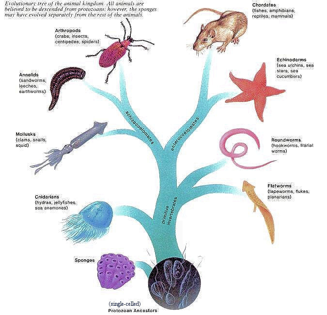
All phyla of animals had evolved by the beginning of the Paleozoic Era some 540 million years ago. The evolutionary tree of animals in Figure 01b indicates that animals are the descendants of protozoans - perhaps in a colonial form whose cells differentiated into various types of cells. The evolutionary tree of animals resembles a tree with 2 main branches. The animal phyla located on the main trunk of the tree are referred to as the primitive invertebrates, and the animals of the main 2 branches include the advanced invertebrates and the vertebrates. Invertebrates lack a dorsal backbone, while vertebrates have a backbone made up of vertebras. A study of the evolution of animals reveals that the most complex animals have the most advanced features as listed in Table 01 below. Classification of animals therefore is based on type of body plan, symmetry, number of |
Figure 01b Evolutionary Tree of Animals [view large image] |
germ layers, level of organization, type of body cavity, and presence or absence of segmentation. |
| Features | Most Primitive | Primitive | Advanced | Most Advanced |
|---|---|---|---|---|
| Body Plan | None | Sac plan | Tube-within-tube plan | + specialization of parts |
| Symmetry | None | Radial | Bilateral | + cephalization§ |
| Germ layers | None | 2 | 3 | 3 |
| Level of organization | None | Tissue | Organ | Organ system |
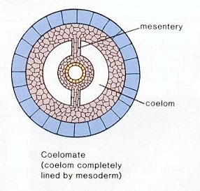
| Body cavity | Diploblastic | Acoelomate | Pseudocoelom | True coelom |
| Segmentation | Nonsegmented | Nonsegmented | Segmented | + specialization of parts |
![]() |
![]() |
necessary for multicellularity already. Their evolutionary steps are clearly demonstrated from single-celled aquatic protists to colonies and then appear as the collar cells in sponges. A sponge does not have anything in their bodies that can be called tissues or organs. Instead each type of sponge cell is responsible for a different activity to keep the sponge alive. Some sponges grow on rocks and are brightly colored. Sponges often are shaped like vases with a central cavity (Figure 02). Figure 03 shows the anatomy of the sponge. The internal structures are described in the followings. |
Figure 02 Sponge |
Figure 03 Sponge Anatomy [view large image] |
Although the terminologies (and hence the functions) for the various systems are in close parallel to those for the human body, the composition and distribution are vastly different. |
![]() |
Germ cells provide the continuity of life between generations. In higher animals (up to roundworms) there is a clear and early separation of germ cells from somatic cell types. For the other species somatic cells can readily become germ cells even in adult organisms such as the buds, and polyps of many invertebrate phyla. For those organisms where there is an established germ line that separates early in development, the germ cells come from the primordial germ cells (PGCs) in the yolk sac and then migrate into the developing gonads (the genital ridges, originally not in the lower part |
Figure 04a Primordial Germ Cells [view large image] |
of the body) as shown in Figure 04a. Once in the gonad, primordial germ cells continue to divide mitotically, producing millions of potential gametes. The PGCs then undergo meiosis to produce the male and female germ cells, i.e., the sperms and eggs. |
![]() |
Fertilization is the process whereby two sex cells (gametes) fuse together to create a new individual with genetic potentials derived from both parents. Although the actual details of fertilization vary enormously from species to species, the events of conception generally consist of four major activities: 1. Contact and recognition between sperm and egg. In most instances, this insures that the sperm and egg are of the same species. |
Figure 04b Sperm and Egg [view large image] |
2. Regulation of sperm entry into the egg. Only one sperm can ultimately fertilize the egg. 3. Fusion of the genetic material of sperm and egg. 4. Activation of egg metabolism to start development. |
![]() |
microtubules. There are three fused microtubules in each group. The two centrioles are arranged such that one is perpendicular to the other. During animal cell division, the centrosome divides and the centrioles replicate. The result is two centrosomes, each with its own pair of centrioles. The two centrosomes move to opposite ends of the nucleus, and from each centrosome, microtubules grow into a "spindle" which is responsible for separating replicated chromosomes into the two daughter cells. It is found that the nucleus of the sperm cell is not necessary for the early divisions at the very beginning of development. It is the centrosome (and the centriole within) that is absolutely essential for the initial divisions. There are cases in which a centrosome can actually be created within the egg. This process is called parthenogeneseis, or virgin birth (see more below) |
Figure 04c Centrosome and Centriole [view large image] |
![]() |
![]() |
Almost all animal species reproduce sexually, which is believed to have evolved as a mean of strengthening the "fitness" of the species by mixing the genes of two different individuals from meiosis. About 1% of animal species reproduce by partenogenesis (Greek for "virgin birth"), while an even smaller fraction switch between sexual and asexual reproduction (known as cyclical parthenogenesis). |
Figure 04d Parthenogenesis Examples [view large image] |
Figure 04e Partheno- genesis [large image] |
Both kinds of parthenogenesis occur most often in low-level species including some fish, amphibians and reptiles (see some examples in Figure 04d). Some experiments try to |
![]() |
![]() |
shape with the mouth region directed downward, are called jellyfishes, or medusae. The polyp is adapted to a sessile life, while the medusa is adapted to a floating or free-swimming existence. At one time, both body forms may have been a part of the life cycle of all cnidarians, because today we see an alternation of generations life cycle of these two forms in certain cnidarians (see Figure 05d). In such life cycle, the polyp stage produces medusae, and the medusae, which produce eggs and |
Figure 05a Cnidaria [view large image] |
Figure 05b Hydra Anatomy [view large image] |
sperm, disperse the species. Cnidarians are quite diversified including the Portuguese man-of-war, sea anemones, hydra and many other species. |
![]() |
![]() |
|
Figure 05c Evolution of Vision [view large image] |
Figure 05d Compound Eye [view large image] |
![]() |
of cells (Figure 05b). For example, they can produce the ovary and the testes and probably account for the animal's great regenerative powers. Like the sponge, a whole cnidarian can grow from a small piece. When conditions are favorable, small outgrowths, or buds, appear, pinch off, and begin to live independently. Figure 05e shows the cnidaria life cycle involving both sexual and asexual reproductions. The larva in the life cycle represents a juvenile form of the animal undergoing metamorphosis (form change). |
Figure 05e Cnidaria Life Cycle [view large image] |
![]() |
Flatworms are nonsegmented, lack a coelom, and have the sac body plan with only one opening. Therefore, if we analyze them according to Table 01, they have a combination of primitive and advanced features. There are three classes of flatworms: one is free living and two are parasitic. The free-living specimen, the planarian, best exemplifies the characteristics of the phylum. Tapeworms and flukes are parasitic with structure reflecting the modifications that occur in parasitic animals. Concomitant with the loss of predation, there is an absence of cephalization; the anterior end notably carries hooks and/or suckers for attachment to the host. The parasite acquires nutrient molecules from the host, and the digestive system is reduced. It is | Figure 06 Tapeworm Life Cycle [view large image] |
covered by a specialized body wall resistant to host digestive juices. The extensive development of the reproductive system, with the production of millions of eggs, may be associated with difficulties in dispersing the species. Figure 06 shows the life cycle of the tapeworm. |
![]() |
|
Figure 07 Planarian Anatomy [view large image] |
eggs hatch in 2-3 weeks as tiny worms. Planarians are capable of regeneration, if a worm is cut crosswise, it usually grows a new head or a new tail as is appropriate. |
![]() |
with the rotifers. Roundworms possess two anatomical features not seen in more primitive animals: a tube-within-a-tube body plan and a body cavity. The body cavity is a pseudocoelom, or a cavity incompletely lined with mesoderm. This fluid-filled pseudocoelom provides space for the development of organs, and serves as a type of skeleton. When roundworms are analyzed according to Table 01, they are seen to have features associated with advanced animals except that they are nonsegmented. |
Figure 08b Roundworm Anatomy |
![]() |
|
Figure 08c Evolution of Mouth and Anus [view large image] |
![]() |
All the advanced invertebrate phyla have a true coelom. Nevertheless, they can be divided into two groups on the basis of embryological evidence (Figure 09). In mollusks, annelids, and arthropods, the coelom forms by splitting of the mesoderm. Therefore, they are called the schizocoelomates. In echinoderms and chordates, the coelom forms by outpocketing of the primitive gut. It is thus called enterocoelomates. Note the interchange of mouth and anus in these two different types of development. |
Figure 09 Coelom Formation |
![]() |
|
Figure 10 Clam Anatomy |
side of the visceral mass, which lies above the foot. Gills are composed of vascularized, highly convoluted, thin-walled tissue specialized for gas exchange. |
![]() |
separatly or in groups under stones, logs or fallen leafs. After a few weeks tiny snails are born. They have a transparent shell. Some snailspecies are ovo-viviparious, this means that the eggs hatch inside of the snail. Most species reach maturity in a year, but the larger ones can take two to four years to reach maturity.
|
Figure 11 Snail Anatomy |
blood and arranged like the teeth of a comb. |
![]() |
|
Figure 12 Digestive System |
ink sac to escape danger. They can also change color according to mood and environ- ment, sometimes exhibiting rapid waves of color changes that sweep over the body. Cuttlefish has internal skeleton; it seems the skin is dotted with light sensors that help to control camouflage. |
![]() |
![]() |
|
Figure 13a Jar-opening Octopus |
Figure 13b Octopus Anatomy |
The octopus anatomy is shown in Figure 13b. |
![]() |
|
Figure 14 Earthworm Anatomy |
efficient absorption of oxygen dissolved efficient in the water. |
![]() |
![]() |
Figure 15B and D) are often fused into regions such as the head, thorax, and abdomen. The head and thorax may be fused to form a cephalothorax (class Crustacea, Figure 15A). Since insects comprise one of the largest animal groups - both in number of species (about 1 million) and in number of individuals, the grasshopper will be used as the anatomical example (Figure 16e), also see a crayfish anatomy in Figure 16b. Figure 16a captures an image of |
Figure 15 Arthropods |
Figure 16a Flight of the Bumblebee [view large image] |
the flight of bumblebee. |
![]() |
|
Figure 16b Crayfish Anatomy |
![]() |
|
Figure 16c Compound Eyes of Mosquito [view large image] |
allow the animal to be active. |
![]() |
of two different ecological settings (niches). For example, caterpillars are specialized for feeding on plant leaves. The butterfly of many species is specialized for finding mates and reproduction.
|
Figure 16e Grasshop -per [view large image] |
![]() |
radially symmetrical, in contrast to vertebrates; they have no internal skeleton, no trace of any of the three major chordate characters of notochord, nerve cord, or gill slits, and they have many peculiar and complicated organs of their own. But the embryology sheds an unexpected gleam of light. The early embryo of the echinoderm is a tiny creature, which floats freely in the sea water. Unlike the adult, the larva is bilaterally symmetrical, suggesting that the radial symmetry of the starfish is a secondary affair, assumed when the ancestors of these forms look up a sedentary existence. Then, too, the type of development of certain of the body cavities is identical with that found in the embryos of some primitive vertebrates. It is believed that the bilateral larva developed types which retained the original symmetry, |
Figure 17 Echinoderms |
and gradually evolved into the chordates and, finally, the true vertebrates. |
![]() |
projecting arms and after some more weeks, a brachiolaria larva is formed. The larvae have their own gut, with inside cilia to inhale and transport food particles. They feed themselves with diatoms and other organisms in the plankton. The stomach is large and round and situated at the backside. After this phase a large part of the larva degenerates and at the rear side a rudimentary formed juvenile starfish develops. The organs of the young starfish are formed anew.
|
Figure 18 Starfish Anatomy |
function of respiratory exchange. |
![]() |
![]() |
the only chordate feature retained by the adult. The larva of the tunicate, however, has a tadpole shape and possesses the three chordate characteristics. It has been suggested that such a larva may have become sexually mature without developing the other adult tunicate characteristics, and may have evolved into a fishlike vertebrate similar to the lancelet, which is a chordate that shows the three chordate characteristics as an adult. |
Figure 19a Vertebrates |
Figure 19b Protochordates |
![]() |
|
Figure 19c Limbs |
sometimes absent). There is a fundamental design in the skeleton of all vertebrates. Humans, bats, lizards, and whales are all just variations on a theme (see for example the common pattern of limbs for some vertebrates in Figure 19c). |
![]() |
Bone is the major component of the skeletons in adult vertebrates. It is composed of both living tissues, such as bone cells, fat cells, and blood vessels, and nonliving materials (such as collagen) secreted by the bone cells called osteoblasts into the inter-cellular space. The collagen fibers are coated with a calcium phosphate salt, making it strong without being brittle. As shown in Figure 19d, a bone can be divided into four parts: |
Figure 19d Bone [view large image] |
1. Periosteum - This fibrous membrane is the outer layer of the bone. It is rich with blood vessels and nerve endings and it ends at the edge of the joint area or where the ligaments and the tendons insert themselves. |
![]() |
An important contribution to the shape of animals with backbones is the number of vertebrae (bones in spinal column) that make up the structure. While human has only 33, snakes have more than 300, with some species having more than 500. Vertebrae develop from segments of tissue called somites, which form, one after another, in a head-to-tail sequence in the embryo (diagram a, Figure 19e). They bud off from the "head" end of the presomitic mesoderm (PSM), an immature tissue fated to generate the somites. This budding is regulated by a "clock-and-wavefront" |
Figure 19e Somitogenesis [view large image] |
model. In snakes, the clock genes seems to express 4 times faster than in shorter-bodies animal (such as mice), leading to many more, though smaller, somites (see diagram b in Figure 19e). |
![]() |
1. Cleavage - a stage of extremely rapid mitotic divisions wherein the zygote cytoplasm is divided into numerous smaller cells. By the end of cleavage these cells generally form a fluid-filled sphere known as blastula. 2. Gastrulation - The cells in the blastula undergo dramatic movements wherein they change their positions relative to one another. As a result, the typical embryo contains three germ layers - the etoderm in the outside, the mesoderm in the middle and the endoderm is the innermost layer. The invagination of cells is something like poking a curved finger into a balloon. 3. Organogenesis - The cells interact with one another and rearrange themselves to produce the bodily organs. In vertebrates, the mid-dorsal ectodermal cells fold to form the neural tube with the notochord laying under. Also during this stage certain |
Figure 19f Embryonic De- velopment [view large image] |
cells such as the blood, lymph, pigment, and the gamete migrate from their place of origin to the final location. 4. Gametogenesis - As shown in Figure 19f, a portion of egg cytoplasm gives rise |
![]() |
![]() |
lungs for gas exchange instead of gills. It is believed that the gill arches have been modified to become the jaw as shown in Figure 20a. Further modification of these gill arches can be traced from an embryo to an adult (in humans as shown in |
Figure 20a Evolution of Jaws [view large image] |
Figure 20b Evolution of Gill Arches [view large image] |
Figure 20b). The origins of jaws, ears, larynx, throat, bones, muscles, nerves, and arteries can all be found in these gill arches. |
![]() |
![]() |
used for respiration. The coelacanth and shark in Figure 21a are the "living fossil" among the fishes. They are species that have defied the evolutionary odds to survive virtually unchanged for tens or hundreds of millions of years. Other examples with such distinction are shown in Figure 21b. |
Figure 21a Fishes |
Figure 21b Living Fossils |
Figure 21a also includes a fish squirting a shot at the cricket. Figure 22 shows the internal anatomy of a common fish. |
![]() |
|
Figure 22 Fish Anatomy |
As the water passes over the gills, oxygen is absorbed by blood and carbon dioxide is given off. |
![]() |
|
Figure 23 Amphibians |
![]() |
Man has 12. Similarly, the frog has only 10 pairs of spinal nerves. Man has 30 pairs. Two simple holes make up the nostrils for the frog. There are complex valves but no long nasal passages as there are in man. The frog's sense of smell is registered by olfactory lobes. These make up the forward portion of the brain. The eye is crude. Its fixed lens cannot change its focus. Poorly developed eyelids do not move. To close its eye, the frog draws the organ into its socket. A third eyelid, or nictitating membrane, may be drawn over the pulled-in eyeball. There is no external ear. Both eardrums, or tympanic membranes, are exposed. There is only one bone in the frog's middle ear. The human middle ear contains three bones (malleus, incus, and stapes in the ossicles). As in man, semicircular canals help to maintain body balance. |
Figure 24a Frog Anatomy |
![]() |
sound-generating mechanisms and organs for acoustic perception. Fishes are able to produce different types of sounds and to perceive acoustic signals of different frequencies, temporal patterns and intensities. They possess inner ear and a few have rudimentary middle ear (the ossicles). Human diving experience indicates that sound wave in water can only be perceived through bone conductivity via vibration of the bones of the skull. The efficiency of this method of sound wave detection is 40% weaker than air conductivity in land animals and it lacks sufficient orientation to identify the direction of the audial source. It is not accidental that the full apparatus of ear developed only after animals making a living on land. Figure 24b traces the development of the stapes from the gill arches in the upper jaw of fish to the stapes in amphibian. The same figure also shows that the malleus and incus come from the lower jawbones of the reptile. The transformation sequence is supported by fossils bearing a continuum of forms as shown. The construction of the mammalian middle |
Figure 24b Ear Bones Evolution [view large image] |
ear allows detection of higher frequency sounds. The distance between the two ears enables its owner to discern direction of the sound source (because the sound speed in air is lower than that in water). See also evolution of the inner ear. |
![]() |
|
Figure 24c Cutaneous Gas Exchange [view large image] |
frog known to science without lungs and joins a short list of amphibians with this unusual trait, including a few species of salamanders and a wormlike creature known as a caecilian. |
![]() |
|
Figure 24c Reptiles |
by exposing themselves to the sun if they need warmth or by hiding in the shadows if they want to cool off. |
![]() |
lizards with a bladder, it is connected to the cloaca by a short urethra. Urine passes into the cloaca and then into the urinary bladder, if present, or into the distal colon where water resorption occurs. The cloaca typically consists of 3 chambers. The coprodeum is the most cranial and receives fecal material and urinary wastes. The urodeum is the middle section and receives genital secretions and urinary wastes |
Figure 24d Lizard Anatomy |
from the urogenital ducts. The caudal proctodeum acts as a reservoir for fecal and urinary wastes before they are excreted. This also is the location of the openings of the musk glands. |
![]() |
fertilization has occurred, the egg is covered by a protective, leathery shell and is laid in an appropriate location. The shelled egg made development on land possible and eliminated the need for a swimming-larva stage during development. It provides the developing embryo with oxygen, food, and water; it removes nitrogenous wastes; and it protects the embryo from drying out and from mechanical injury. This is accomplished by the presence of extraembryonic membranes (Figure 24e). |
Figure 24e Membranes |
![]() |
Birds are characterized by the presence of feathers, which are actually modified reptilian scales. There are many orders of birds, including birds that are flightless (ostrich), web footed (penguin), divers (loons), fish eaters (pelicans), waders (flamingos), broad billed (ducks), birds of prey (hawks), vegetarians (fowl), shorebirds (sandpipers), nocturnal (owl), small (hummingbirds), and songbirds, the most familiar of the birds. Some of them are showed in Figure 25a. |
Figure 25a Birds |
Nearly every anatomical feature of a bird can be related to its ability to fly. Figure 26a shows the anatomy of a common bird. |
![]() |
|
Figure 25b Warm Blooded- |
shows the temperature difference between the warm-blooded human and cold blooded lizard, it also shows the temperature differences from different parts of the body. The 37 oC is measured in the mouth, it is 37.4 oC in the rectum, and 36.7 oC in the armpit. |
![]() |
![]() |
![]() |
feathers and can walk/swim (ducks); while others incubate eggs a short time - chicks are born featherless and helpless (robins). The process of making egg is illustrated in Figure 26b; while the structure of a chicken egg is |
Figure 26a Bird Anatomy |
Figure 26b Making Egg |
Figure 26c Chicken Egg |
displayed in Figure 26c together with a description of its various components in Table 02. |
| Component | Composition | Function(s) |
|---|---|---|
| Yolk | Protein and fat | Provision of nourishment |
| Albumen | Protein and water | Provide additional nutrition and protection |
| Amniotic fluid | Water and other materials | Protection and aiding the growth of the fetus |
| Allantois | Nitrogenous waste | Collection of waste |
| Embryonic disk | Cells of the embryo | The developing embryo |
| Chalaza | Part of egg white, 2 spiral bands of tissue | Stabilizing the yolk |
| Chorion | Two layers of cells | Membranes for gas exchange |
| Air sac | Air | Provide oxygen for baby chick just before hatching |
| Shell | Protein matrix lined with calcium carbonate | Protective outer cover |
![]() |
The chief characteristics of mammals are hair and mammary glands that produce milk to nourish the young. Human mammary glands are called breasts. Mammals are classified according to their means of reproduction: there are egg-laying mammals called monotremes such as the duck-billed platypus; mammals with pouches for immature embryos are the marsupials such as the kangaroos; while the placental mammals are the majority of living mammals. Figure 27a shows just a few of these animals. Table 03 lists the 18 orders of living placental mammals. They are classified largely according to the mode of locomotion and how they get their food. Figure 28 illustrates a cat's anatomy, |
Figure 27a Mammals |
which is very similar to the human's. Information on "mammal characteristics and comparison with other vertebrates" can be found in the appendix on "Age of Animals". |
| Order | Examples | Characteristics |
|---|---|---|
| Edentata (Xenarthra) |
Anteater, armadillo, sloth | Primitive terrestrial mammal; few or no teeth; well developed claws |
| Pholidota | Pangolin | Medium size; large, plate-like scales; lack teeth, use powerful front claws and long tongues to reach ants or termite |
| Lagomorpha | Rabbit, hare, pika | Chisel-like incisors; hind legs longer than front legs; herbivorous |
| Rodentia | Mouse, rat, squirrel, beaver, porcupine | Incisor teeth grow continuously |
| Macroscelidea | Elephant Shrew | African species of shrew-like creatures with long nose |
| Primates | Lemur, monkey, gibbon, chimpanzee, gorilla | Mostly tree dwelling; head freely movable on neck; 5 digits, usually with nails; thumbs and/or large toes usually opposable |
| Dermoptera | Flying Lemur | With "wings of skin" to support gliding, under-developed new born |
| Scandentia | Tree Shrew | High brain/body mass ratio, live in trees, under-developed new born |
| Chiroptera | Bat | Digits support membranous wings |
| (Eu)lipotyphia (Insectivora) |
Mole, shrew | Primitive; small, sharp-pointed teeth |
| Carnivora | Dog, bear, cat, sea lion | Long canine teeth; pointed teeth |
| Artiodactyla | Pig, camel, buffalo, giraffe | Medium to large; 2/4 toes, each with hoof; many with antlers/horns |
| Cetacea | Whale, porpoise | Medium to very large; paddlelike forelimbs; hind limbs absent |
| Tubulidentata | Aardvark | Pig-like animal with powerful claws & long tongue for eating termites |
| Perissodactyla | Horse, zebra, tapir, rhinocerose | Large, long-legged, one or 3 toes, each with hoof; grinding teeth |
| Hyracoidea | Hyrax | Plant-eating with short ears and has toenails resembling hooves |
| Proboscidea | Elephant | Large size with trunk, pillow-like limbs, broad and padded foot |
| Sirenia | Manatee | Slow moving aquatic mammals with flippers, flattened tail, no legs |
![]() |
|
Figure 27b Brains of the Vertebrates |
amphibian, a reptile, a bird, and a mammal. See also layers of the reptilian, mammalian, and human brains. |
![]() |
![]() |
appearance of the extraembryonic membranes and the formation of the umbilical cord in the human embryo. The embryonic disk had initially pressed to the wall of the uterus at implantation. It had moved around during the development for attaching the umbilical cord in the tummy |
Figure 27c Early Embryo [large image] |
Figure 27d Fertilization - Implantation |
button to the placenta. Figure 27d depicts the progression from fertilization to implan-tation in the earliest stage of development. |
![]() |
contract to some degree. As a result, air is sucked into and pushed out of the lungs through the trachea and the bronchial tubes or bronchi; these branch out and end in alveoli which are tiny sacs surrounded by capillaries filled with blood. Here oxygen from the air diffuses into the blood, where it is carried by hemoglobin. The deoxygenated blood from the heart reaches the lungs via the pulmonary artery and, after having been oxygenated, returns via the pulmonary veins. |
Figure 28 Cat Anatomy |
![]() |
Humans are mammals in the order Primates. The first primates may have resembled today's tree shrews, rat-size animals with a snout, claws, and sharp front teeth. By 50 million years ago, however, primates had evolved characteristics suitable to move freely through the trees. The first primates were prosimians (meaning "premonkeys"). They are represented today by several types of animals, including the lemurs. Monkeys, along with apes and humans, are anthropoids. Monkeys evolved from the prosimians about 38 million years ago, when the weather was warm and vegetation was like that of a tropical rain forest. There are two types of monkeys: the New World (South America) monkeys such as the spider monkeys, which have long grasping tails and flat noses, and the Old World (Africa) monkeys such as the baboons, which are now ground dwellers and lack such tails. Ape (gibbon, gorilla, and chimpanzee) evolved later. The human lineage split from that of the apes occurred about 5 - 10 million years ago in Africa. Figure 29 shows some of the primates. Figure 30 illustrates the chimpanzee anatomy, which is virtually identical to the human's. |
Figure 29 Primates |
![]() |
living. It is useful as an effective feeling and grasping mechanism to grab their insect prey, and to prevent them from falling and tumbling while moving through the trees. By far the most outstanding characteristic of primate evolution has been the enlargement of the brain among members of the order. Primate brains tend to be large, heavy in proportion to body weight, and very complex.
|
Figure 30 Chimp Anatomy |