Mulitcellular Organisms
Sleep and Dream
 |
 |
Sleep is another process in the brain that we still have much to learn. We know that while we sleep, we cycle between two very different states. The first is called slow wave sleep characterized by long waves of undulating electrical activity. The second is rapid eye movement (REM) sleep characterized by frantic brain activity that looks very much like wakefulness. It also has very obvious physical signs: the rapid flickering motion of the eyeballs, the near-total
|
|
|
muscle paralysis (to prevent from acting out the dreams), and penile erections.
|
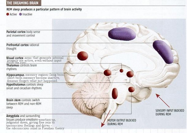
It is found that dreams are associated with REM sleep. During dreaming, the visual cortex is very active (to generate internal imagery), as are the amygdala, thalamus and the brainstem, which fits with the fact that dreams tend to be very visual and emotional. At the same time, the prefrontal and parietal cortices and the posterior cingulate, areas which deal with rational thought and attention, are all very quiet, which tallies with the lack of insight, illogicality and time distortion that characterizes dreams. Although the hippocampus is actively processing long-term memory, the short-term memory region is inactive, which explains why dreamer forgets what just happened (see Figure 10-18).
It is now known that REM sleep falls into two types, generating two different kinds of dreams. Firstly there is the tonic component. It is accompanied by muscle relaxation and sometimes sexual arousal. Tonic REM takes place earlier on in the sleeping cycle. It is calmer, more restful, and more passive. When woken, the dreamer typically reports such things as "I was
 |
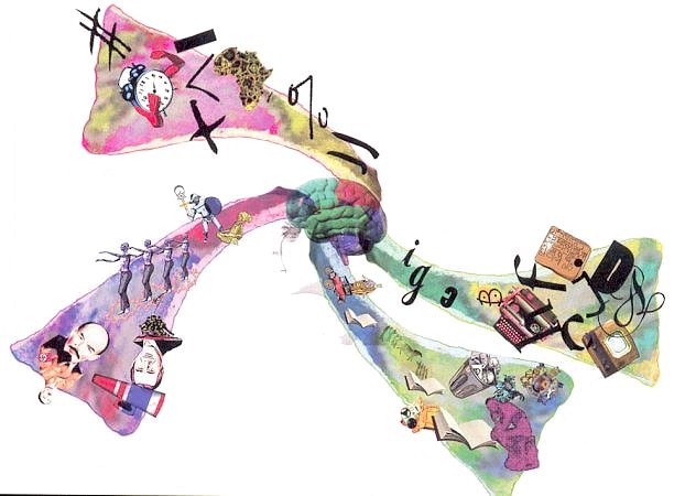 |
feeling floaty" (Figure 10-19) or "there was a feeling of peace". The second type of REM sleep is known as phasic and is characterized by jerky eye movements, spasmodic limb and facial twitching and sudden breathing changes. When volunteers are woken from this sort of REM sleep, they typically describe their dreams as being strongly visual, active and "real". Phasic REM and its accompanying dreams tend to occur later on in the sleeping period. Nightmares are associated with this type of
|
|
|
sleep (see Figure 10-20a).
|
 |
REM sleep appears to have arisen quite early in evolution - worms, insects, reptiles, birds and mammals all do it. Therefore, it must serve a very useful function. There are many theories of sleep function, which fall into four broad classes: restoration and recovery, predator avoidance, energy conservation and information relocation from short-term to long-term memory, (discarding redundant data in the process, Figure 10-20b) similar to the transfer of data from
|
|
disk to tape in the IT (information technology) industry. But not one of them has been confirmed or refuted. Figaure 10-21 shows the sleep patterns (or the lack of it) for a variety of species.
|
Recent research in 2003 indicates that non-REM sleep may give brain cells a chance to repair themselves, and REM sleep may allow the brain's neuron receptors to recover (regain full sensitivity). It is found that Animals born inmature require more REM sleep. Thus REM sleep may also act as a substitute for the external stimulation that prompts neuronal development in creatures that are mature at brith. Sleep research will identify the brain regions that control REM and non-REM sleep. It will lead to a more comprehensive and satisfying understanding of sleep, its functions, the mechanisms and evolution. It will probably gain insights into exactly what is repaired and rested, why these processes are best done in sleep.
It is now realized that sleep is an actively regulated process, not simply the passive result of diminished waking, and that sleep should be regarded as a reorganization of neuronal activity rather an cessation of activity. It is found that the vigorous brain
 |
 |
activation of REM sleep occurred at regular 90-minute intervals and occupied up to 20% of sleep. Even during NREM sleep, when consciousness may be totally obliterated, the brain remains significantly active. There is only a 20% reduction in cerebral blood flow during sleep. Although consciousness is dulled, the brain is still roughly 80% activated and thus capable of robust and elaborate information processing. It is amusing that all these activities occur inside our brain every night
|
|
|
but we have only a dim notion of what is going on.
|
Figure 10-22a summarizes the sleep states.
- A "Wake" state including stages 1 and 2 has been added in the Diagram. The actual sleep cycle involves only the NREM (in brown color), and the REM (in green).
- Behaviour - Changes in position can occur during waking and in concert with phase changes of the sleep cycle. Two different mechanisms account for sleep immobility. The first is disfacilitation (during stages 1- 4 of NREM sleep). The second is inhibition (during REM sleep). During dreams, we imagine that we move, but we do not.
- Awake - Waking (in the present context) is the phase during which the body prepares for sleep. All people fall asleep with tense muscles, their eyes moving erratically. Then, normally, as a person becomes sleepier, the body begins to slow down. Muscles begin to relax, and eye movement slows to a roll.
- Sleep can be divided into five stages. Although the signals for transition between the five stages of sleep are mysterious, it is important to notice that these stages are discretely independent of one another, each marked by subtle changes in bodily function and each part of a predictable cycle whose intervals are observable. Sleep stages are monitored and examined clinically with polysomnography, which provides data regarding electrical states of the muscle (electromyo-gram - EMG), the brain (electroencephalogram - EEG), and eye movement (electrooculogram - EOG) during sleep.
- Stage 1 - This is a stage of drowsiness. Polysomnography shows a 50% reduction in activity between wakefulness and stage 1 sleep. The eyes are closed during Stage 1 sleep, but if aroused from it, a person may feel as if he or she has not slept. Stage 1 may last for five to 15 minutes.
- Stage 2 - Stage 2 is a period of light sleep during which polysomnographic readings show random fluctuation. These waves indicate spontaneous periods of muscle tone mixed with periods of muscle relaxation. The heart rate slows down, and body temperature becomes lower. At this point, the body prepares to enter deep sleep.
- Stage 3 - This stage is known as slow-wave, or delta, sleep. During slow-wave sleep the EMG records slow waves of relatively high amplitude, indicating a pattern of deep sleep and rhythmic continuity.
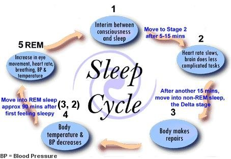
- Stage 4 - This stage is similar to Stage 3 but more intense. The period of non-REM sleep (NREM) is comprised of stages 1 - 4 and lasts from 90 to 120 minutes. In addition, stage 2 and 3 repeat backwards before REM sleep is attained. Therefore, a normal sleep cycle has this pattern: stage 1, 2, 3, 4, 3, 2, REM. Usually, REM sleep occurs 90 minutes after the onset of sleep.
- REM - REM sleep is distinguishable from NREM sleep by changes in physiological states, including its characteristic rapid eye movements. However, polysomnograms show wave patterns in REM to be similar to Stage 1 sleep. In REM sleep, heart rate and respiration speed up and become erratic, while the face, fingers, and legs may twitch. Intense dreaming occurs during REM sleep as a result of heightened cerebral activity, but paralysis occurs simultaneously in the major voluntary muscle groups. Because REM is a mixture of encephalic (brain) states of excitement and muscular immobility, it is sometimes called paradoxical sleep. The first period of REM typically lasts 10 minutes, with each recurring REM stage lengthening, and the final one lasting an hour. The percentage of REM sleep is highest during infancy and early childhood, drops off during adolescence and young adulthood, and decreases further in older age.
A sleep cycle (see Figure 10-22b) comprises five stages of sleep, including their repetition. The first cycle, which ends after the completion of the first REM stage, usually lasts for 100 minutes. Each subsequent cycle lasts longer, as its respective REM stage extends. So a person may complete five cycles in a typical night's sleep.
- Sample tracings of three variables used to distinguish the state are also shown in Figure 10-22a: The EMG tracings are highest during waking, intermediate during NREM sleep and lowest during REM sleep. The EEG and EOG are both activated during waking and REM, but inactivated during NREM sleep. Each sample shown is approximately 20 seconds long.
- The three bottom rows in Figure 10-22a describe other subjective and objective state variables such as sensation, perception, thought, and movement.
There are essentially two theories on the requirement of sleep - recovery from exertion and consolidation of memory. A 2010 experiment on zebrafish synapses reveals that it has lower overall synapse activity during sleep. It shows that sleep is a process to reduce the activity in the brain preventing overload. However it is also found that not all neural circuits are affected by sleep in the same way with learning and memory reaping the most benefit. Thus the two hypotheses about sleep may not be mutually exclusive.
 |
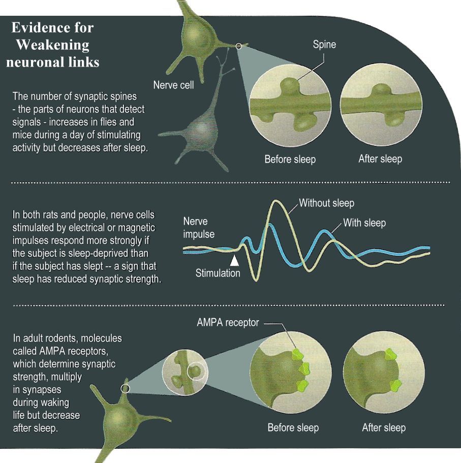 |
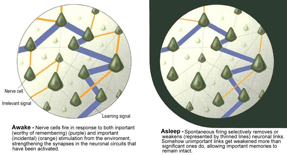
An article in the August 2013 issue of Scientific American indicates that instead of augmenting the neural connections to preserve the memory, the links are actually pruned during sleeping (Figure 10-23a). Presumably, the process gets rid of those un-important events in order to conserve space, energy, and to reduce stress on the nerve cells. Such weakening would return the synapeses to a baseline level of strength. Some of the evidences obtained
|
Figure 10-23a Sleep and Neuron Pruning [view large image] |
|
from flies and mice (and human too) are shown in Figure 10-23b.
|
.


