Home Page Overview Site Map Index Appendix Illustration About Contact Update FAQ


Mitosis and Meiosis


Contents

DNA Organization
Mitosis
Meiosis

DNA Organization

    Most DNA resides within a region called nucleolus in eukaryotic cell and nucleoid in prokaryotic organism. Extrachromosomal DNA can be found in organelles such as mitochondria, chloroplast or plasmid - all in the shape of a ring. The DNA in the nucleolus is organized in various forms depending on the cell cycle. It is portrayed pictorially in Figures 01, and 02 and described briefly in the followings.

  1. Nucleosome - The basic unit of DNA packing in eukaryotes is the nucleosome, which consists of a segment of DNA wrapping around four pairs of histone protein cores (upper left insert in Figure 01). About 200 cm of linear DNA have to be packed into a core of roughly 10-4 cm diameter. The core particles are connected by stretches of "linker DNA" Linker histones such as H1 are involved in chromatin compaction and sit at the base near the DNA entry and exit points.

  2. Chromatin - This structure is created by linking the nucleosomes together (Figure 01). Its functions are : (a) to package DNA into a smaller volume, (b) to provide an environment for mitosis, (c) to prevent damage to DNA, and (d) to to control gene expression and DNA replication. It is found only in eukaryotic cells and normally folded under the nuclear envelope. It exists in two different states :
  3. Chromosome - This is the even more tightly packaged form of chromatin. While the DNA and RNA within are information-carrying macromolecule, the proteins serve to package the whole thing and control its functions. The present of such form is a signal for cell division in either mitosis or meiosis. Initially, it is a single-chromatid (single stranded) chromosome; it soon replicates itself to form a double stranded chromosome (as shown in most text books and science magazines, see all the forms in Fgiure 02).
Nucleosome Chromatin

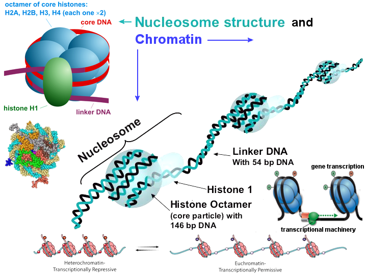
Figure 01 Nucleosome
[view large image]


Figure 02 Nucleosome, Chromatin, and Chromosome [view large image]


[Top]


Mitosis

Mitosis involves the division of the mother cell into two daughter cells, genetically identical to each other and to their parent cell. Mitosis occurs only in eukaryotic cells; while animals undergo "open" mitosis, where the nuclear envelope breaks down before the chromosomes
Cell Cycle Time Cell Cycle Samples 
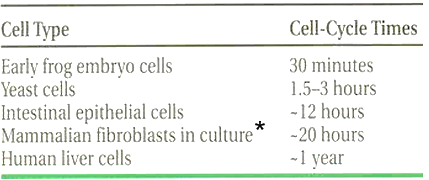
separate, fungi undergo a "closed" mitosis, where chromosomes divide within an intact cell nucleus. Prokaryotic cells, which lack a nucleus, divide by a different process called binary fission. In cultured human cells (among the list in Table 01), about 80% of the cell cycle (~ 24 hrs, see Figure 03) are spent in growth and synthesis. The rest can be categorized into six stages as shown pictoraly in Figure 04, summarized in Table 02, and elaborated further below.
Figure 03 Cell Cycle (also see aging of organs) Table 01 Cell Cycle Samples, * fibroblast is a type of cell that synthesizes the extracellular matrix and collagen, the structural framework for animal tissues, and plays a critical role in wound healing.
  1. Interphase - This is the phase when the cell grows in size and synthesizes mRNA and proteins in preparation for subsequent steps leading to mitosis. In human somatic cells, the interphase takes up about 15% of the cycle time.

  2. Prophase - This process involves condensation of the chromatid into double strands chromosomes. Since the genetic material has been replicated in the prior interphase of the cell cycle, there are two identical copies of each chromosome in the cell. Those copies are called sister chromatids and they are attached to each other at the centromere, which is the center for the microtubules to split the cell in animals.

  3. Prometaphase - In this phase the nuclear membrane breaks apart into numerous "membrane vesicles". The chrommosomes start to interact with the spindle emanated from the centrosomes at the poles.

  4. Metaphase - This is the stage in which the chromosomes align in the equator of cell preparing for the process of separation. The centromeres all line up at equidistance from the two centrosome poles.

  5. Anaphase - In this phase, the chromosomes are split and the sister chromatids move to opposite poles of the cell. The reaction force of pulling begins to stretches the cell into an oval shape.

  6. Telophase - This is the final stage when a filament ring contracts to cleave the mother cell into two daughter cells. The chromosomes are uncoiled from the spindle fibres, and unwind back into the expanded chromatin that is present during interphase.

Mitosis
# Stage Events
1 Interphase Preparing for cell division
2 Prophase Duplication to two sister chromatids
3 Prometaphase Nuclear membrane disintegrated
4 Metaphase Duplicated chromosomes at equator
5 Anaphase Sister chromatids separated
6 Telophase Diploid chromosomes established at each pole, the cell divided into two daughter cells

Figure 04 Mitosis [view large image]

Table 02 Stages in Mitosis


[Top]


Meiosis

Meiosis is a special kind of cell division that produces gametes (sperm and egg). Prior to the initiation of this process, the genetic materials is in the form of indistinct threads called chromatins (see Figure 01), which condense to chromosomes at the time of cell division. At the beginning of meiosis in a cell of the ovary or testis, the randomly distributed chromosomes (from each parent) pair up, side by side. Each chromosome pair then copies itself exactly to make four strands linked by a centromere, which is shown in the diagram as homologous pair.

Meiosis

At crossover points, sections of a chromosome pair are cut and swapped. The crossover points can vary in each of the four strands. Once the genetic material is shuffled, pairs of chromosomes, now with new gene combinations, are pulled into two new nuclei as the cell divides into two (meiosis I). The gametes are produced by one more division (meiosis II). Each of the gamete contains only one copy of the newly shuffled gene.

Genetic variability depends on the different alignment of the chromosome pairs during the meiosis I division. The two newly shuffled versions can place themselves on either one of the daughter cells. There are 23 chromosome pairs in human cell, therefore they can line up in 223 = 8,388,608 different ways. Any one of a person's more than 8 million possible combinations of chromosomes can combine with any one of the more than 8 million combinations of his or her partner, raising potential variability to more than 70 trillion (8,388,6082) genetically unique individuals. Crossover (sometimes referred to as recombination) contributes even more variation.

Figure 05 Meiosis
[view large image]


In short, the differences between mitosis and meiosis are : meiosis has a recombination phase and the cell divides twice to attain a haploid state.