Home Page Overview Site Map Index Appendix Illustration About Contact Update FAQ


Slime Moulds Life Cycle

Multicelluarity 
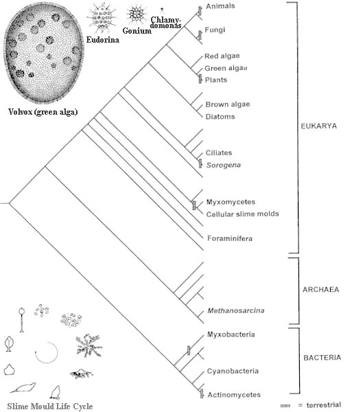
Slime moulds inhabit forest soil and consume bacteria and yeast, which they track by chemotaxis - chemical gradient sensing (of folic-acid in this case). Starvation, however, prompts the normally unicellular cells to aggregate and develop to a true multicellular organism, producing a fruiting body comprised of cellular, cellulosic stalk supporting a bolus of spores. Thus, slime mould has evolved mechanisms that direct the differentiation of a homogeneous population of cells into distinct cell types, regulate the proportions between tissues and orchestrate the construction of an effective structure of the dispersal of spores. Many of the genes necessary for these processes in slime mould were inherited by Metazoa (animal with specialized cells) and fashioned through evolution for use within many different modes of development. Analysis of the proteins shows that slime mould diverged from the animal-fungal lineage after the plant-animal split, but it seems to have retained more of the diversity of the ancestral genome than have plants, animals or fungi, i.e., it possesses a level of complexity that is greater than the yeast, but much simpler than plants or animals. It represents one of the earliest branches from the last common ancestor of all eukaryotes, it is also one of the 13 separate inventions of multicelluarity (Figure 01). Insert in the same image shows the relatives of green algae going from the single cell Chlamydomonas to the 16-cell Gonium, Eudorina, and finally to the largest Volvox, which may consist of 50000 or more cells.

Figure 01 Multicelluarity
[view large image]

Slime Mould 1
    The slime mould life cycle (Figure 02), which alternates between a unicellular feeding stage and a multicellular reproductive stage is described in the followings:

  • They begin their life from spore germination and multiplication.
  • The basic units of slime moulds are haploid amoebae, usually prowling around on the forest floor consuming bacteria.
  • Once the supply of food is exhausted, individual amoebae begin to move together. They form streams of cells called pseudoplasmodium moving slowly at about one millimeter/hour. It is now known that the process is initiated with the cAMP1 signaling molecules released by the starving amoebae.
  • Eventually the streams come together and form an aggregation which is at the spot with high concentration of cAMP. The mass can number a hundred thousand cells and might reach a size of a few millimeters.
  • Then they stick together by secreting adhesion molecules and creating a slime sheath (cap) which covers all the cells in the mass. The polarity defined by the anterior (front), posterior (back) ends is established by oxygen gradient. The responsible gene for adhesion can be identified by blocking its expression. The resulting mass becomes a loose rubble that is incapable of further development.

Figure 02 Slime Mould Life Cycle [view large image]

Slime Mould 2
  • The mass of cells now begins to behave as one, gliding around on the ground as if it were a miniature garden slug leaving a track of slime behind (and hence the name, Figure 03). The movement of the slug is guided by the higher concentration of cAMP at the anterior region of the cap.

Figure 03 Multicelluar Development [view large image]

  • The migration continues toward higher concentration of ammonia (giving off by decaying organism), which means food for them.
  • Slime Mould Video

          Slime Mould in Action
                 [View Video]

    The news about strange organism in 1973 has been largely forgotten for about 46 years until 2019 when the blob goes on public display at the Paris Zoological Park, as part of an exhibition to showcase its rare abilities. See the news and video below, courtesy of CNN.
    Slime Mould

     [See CNN News on the Blob]

    1cAMP is one of the morphogens (signaling molecules) identified in slime mould. The others include ammonia, DIF (signal to induce stalk formation), adenosine, and calcium.