Home Page Overview Site Map Index Appendix Illustration About Contact Update FAQ


Evolution of Micro-organisms and Plants


Contents

Pre-Cambrian Era, 4570 - 541 MYA (Updated 2022)
Cambrian Period, 541 - 485.4 MYA
Ordovician Period, 485.4 - 443.4 MYA
Silurian Period, 443.4 - 419.2 MYA
Devonian Period, 419.2 - 358.9 MYA
Carboniferous Period, 358.9 - 298.9 MYA
Permian Period, 298.9 - 252.2 MYA
Triassic Period, 252.2 - 201.3 MYA
Jurassic Period, 201.3 - 145.0 MYA
Cretaceous Period, 145.0 - 66.0 MYA
Tertiary Period, 66.0 - 2.588 MYA
Quaternary Period, 2.588 MYA - present

The Geologic time scale is adopted from the 30 December 2011 version by the International Union of Geological Sciences (IUGS). MYA = million years ago, FA = first appearance.


Pre-Cambrian Era, 4570 - 541 MYA (Updated 2022)

Pre-Cambrian Family Tree At the time of the appearance of the first organisms about 3.8 billion years ago (see Figure 11-04a), there was no free oxygen, as there is now, but rather a "reducing atmosphere" composed of methane, carbon dioxide, ammonia, and hydrogen (see Figure 11-02a). The microorganisms of this period utilized methane or hydrogen rather than oxygen in their metabolism - they are therefore referred to as "anaerobic" (non-oxygen-using). Fermentation is modern example of anaerobic metabolism. This type of metabolism is 30 to 50 times less effective than oxygen-based ("aerobic") metabolism, or respiration.

Figure 01 Pre-Cambrian Era [view large image]


Figure 02 Family Tree


Mitochondria Chloroplasts Up to 700 MYA, life remained fairly primitive, the distinctions between plants and animals were not very clear-cut. For example, the bacteria (monera) show a variety of forms. Some take in chemicals and depend on atmospheric CO2, like plants (autotrophs - from Greek autos = self, trophe = nourishment). Others take in organic material for food, like animals (heterotrophs - from Greek heteros = other). Some autotrophic bacteria, called chemosynthetic, do not need light; they obtain their

Figure 03 Mitochondria


Figure 04 Chloroplast [view large image]


energy from mineral chemical reactions, such as the conversion of sulfur to sulfate or the production of CH4 from CO2 and H2.


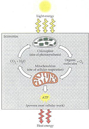
Ecocycle Among the multitude of micro-organisms, two bacteria destined to become the major components for the further development of the living world. The mitochondria (see Figure 03) are the sites of the principal oxidation reaction linked to the assembly of ATP, which supplies energy to most of the organisms today. Their closest relative among present-day bacteria have been identified to be the alpha-proteobacteria. The chloroplasts (see Figure 04) are the agents of photosynthesis in unicellular algae and plants. They store the solar energy in the form of glucose (sugar), which becomes food for the "heterotrophs". Their present-day relative are the cyanobacteria (formerly known as blue-green algae), which is believed to be responsible for the first generation of atmospheric oxygen. As shown in Figure 05a the chloroplasts supply food and oxygen for the heterotropic organims, which in turn produced CO2 for photosynthesis. This ecocycle generates a lot of atmospheric oxygen as shown in Figure 11-02 today. The evolutionary steps for the micro-organisms are shown in Figure 01, which identifies the first organisms to be anaerobic (anoxygenic) autotrophy.

Figure 05a Ecocycle

It is now recognized that the intermediate between bacteria and eukaryotes are the archaea. These organisms have become the focus in the study on the evolution of eukaryotic cells. A living descend from the ancient lineage of archaea called Asgard Archaea has finally been cultured successfully in 2020 after 12 years' effort (see "Meet the relatives of our cellular ancestor"). The resulting cells are tiny spheres 300–750 nanometres in diameter, but they often extrude longer, branched filaments that reach out to meet neighboring bacteria
Asgard Archaea (Figure 05b). The researchers think that such a partnership, both biochemical and physical, could tell us more about the processes that led to the eukaryote cell being formed. These living archaea set the stage for the use of molecular and imaging techniques to further elucidate the metabolism of the archaea and the role of ESPs (Eukaryotic Signature Proteins usually found only in eukaryotes) in archaeal cell biology. This, in turn, could guide the direction of future work investigating how eukaryotic cells emerged.

Figure 05b Asgard Archaea
[view large image]

They also have to found out how did the archaea acquire a nucleus to become eukaryotic.

Updates from further researches :
[2022 Update]

By 2022, scientists are able to examine a few proteins from the archaea - H. volcanii (H) and S. acidocaldarius (S) (see Table 01).
Archaea Proteins These proteins are also found in eukaryotic cells, performing different functions. It indicates that these microbes are related to each others although the original purpose has been lost by evolution in billion years. The genes for these proteins actually exist across a variety of microbes and can be traced all the way back to the "Last Universal Common Ancestor (LUCA)' of all living cells .

Table 01 Archaea Proteins
[view large image]

In 1984, when there was not much knowledge about archaea, a novel hypothesis about the merger of mitochondria to archaea was proposed. It begins with archaea and bacteria (mitochondria) hanging out, sharing resources. The archaeon might start to stretch and bulge its exterior membranes to boost the surface area for nutrient exchange. With time, those bulges might spread and grow around the bacteria until the bacteria were, more or less, inside the archaeon. At the same time, the archaeon’s original exterior membrane, now dwarfed by the long tentacles surrounding it, would evolve into the boundary of the new nucleus, while the cell’s new exterior membrane would form when some particularly long tentacles grew right around the edge, greatly enlarging the cell compared to its archaeal precursor. This process differs from phagocytosis, in that it starts with a community of organisms and takes place over long timescales, rather than in a single bite (aee "The mysterious microbes that gave rise to complex life").

Archaea Proteins Phagocytosis plays an essential role in the older models which have been around since 1967 (aee "Evolutionary Origin of Mitochondria"). Such hypotheses assume that the cells which eventually became eukaryotic were already quite complex, with flexible membranes and internal compartments, before they ever met the bacterium that was to become the mitochondrion. These theories require cells to have developed a way of gobbling up external material, known as phagocytosis, so they could snap up the passing bacterium in a fateful bite (see Figure 05c to compare the difference between the 2 hypotheses).

Figure 05c [view large image] Theories of Mitochondria Merger

As shown in the last row of Table 01, the bacteria and eukaryotes have the same kind of membrane. This feature cannot be explained by the phagocytosis models, in which the eukaryote would have its own membrane long before the merger.
[End of 2022 Update]

Blue-green Algae Another hypothesis proposes that the first organisms were "heterotrophs"; they derived their food from other organisms or organic matter which they were able to consume. Very soon all the available organic matter was exhausted, and life would have cannibalized itself to extinction, were it not for the appearance of a new type of organisms, capable of manufacturing their own food. They are the very early green plants, which were actually an extremely primitive form of algae, similar to the modern blue-green algae, which assume many forms from one-celled to colonial or filamentous (loose association of cells, Figure 06a). But the generation of oxygen in the atmosphere started a crisis for the anaerobic organisms, which became either extinct or have to adopt to the new environment. Since then most of the organisms acquire their energy from aerobic metabolism. The organisms evolved into animals captured mitochondria for this purpose. While the plant cells incorporated both mitochondria and chloroplasts to survive. They still retain copy of their own DNA within. The DNAs are in a circular form reflecting their more primitive origin (from the bacteria).

Figure 06a Blue-green Alage

See more about Mitochondria here.

Sulfur Spring Photosynthesis Fossil evidences have been accumulated over the years that anoxygenic photosynthesis were used by bacteria 3.4 billion years ago in a sulfuric environment such as the hot spring shown in Figure 06b. It is also known that oxygen-producing form of photosynthesis emerged about one billion years later. The question is : why it takes so long ?

Figure 06b Sulfur Spring [view large image]


Figure 06c Photosynthesis Evolution
[view large image]


    The following evolutionary sequence is a probable scenario to explain the train of events that leads to the present day photosynthesis.

  1. Green Sulfur Bacteria - Hydrogen sulfide H2S is a close relative of water H2O. They belong to the same group in the periodic table except that the electron configuration of H2S is further away from the nucleus. Such structure makes H2S easier to break into S + 2H+ + 2e-. The conventional view is for the photosynthesising bacteria first exploited the easy-to-oxidise molecules as shown in Figure 06c (1) in the type-I reaction similar to the photosystem I in Figure 04 with H2O replaced by H2S (see examples of oxidation in Redox Gradients).

  2. Proto-Cyanobacteria (H2S) - Early in their history the green sulfur bacteria experienced some kind of genetic glitch which duplicated the entire set of genes for the type-I reaction. This spare copy was free to modify the codes (without harming its host). Eventually, it evolved an ability to recycle electrons but can only make the ATP molecule to store energy, it could also produce oxygen (Figure 06c (2)). This is called type-II reaction similar to the photosystem II in Figure 04 with H2O replaced by H2S. Thus, this kind of proto-cyanobacteria contained both the type-I and type-II reaction centers. However, they were not linked, only one type could be turned on at a time. The type-II reaction would be switched on when the H2S concentration ran low. This theory will be vindicated if such "missing link" bacteria with no coupling between the two types have ever been found.

  3. Proto-Cyanobacteria (Mn) - It happened on occasions that the proto-cyanobacteria got swept into shallow ponds with no H2S but rich in manganese (Mn). They could duly switch on the type-II reaction to get the electron supply from Mn. However, it didn't help because the manganese produced too many electrons choking up the organisms (Figure 06c (3)).

  4. Cyanobacteria - Until one day the type-I and type-II centers were turned on and coupled, then the excessive electrons could go over to the type-I reaction to relieve the blockage. The modern version of photosynthesis were in place with the manganese getting the electrons from H2O (Figure 06c (4)). Since this kind of modification involving two coincidences (the use of Mn and the link between the two types) is highly improbable, therefore it took a long time (about 1 billion years) to institute.
There is a contentious scenario in which the type-II center evolved first. It was acquired by another group and gradually being modified to the type-I center including the manganese cluster which generates oxygen. These two types merged later via gene transfer. According to this theory, the "missing link" is the bacteria with oxygen production capacity type-I center only (nicknamed indigo bacteria). The debate between these two hypotheses can be settled by the discovery of living representatives of either the proto-cyanobacteria or the indigo bacteria. Whatever the true sequence of photosynthesis evolution, the outcome is hugely important to the world we are inherited.

Stromatolites Not all of the single-celled organisms of this time were solitary. Beginning around 2.4 billion years ago blue-green algae (cyanobacteria) would often grow into large mats, called Stromatolites (see Figure 07). Modern-day Stromatolites can still be found in a sheltered bay (Shark Bay) in West Australia, where the water is so salty that creatures that would otherwise eat them are not able to exist.

Figure 07 Stromatolites


Green Algae Plant Cell Then evolution took another step toward complexity about 1.5 billion years ago. The bacteria acquired a nucleus and advanced to eukaryotic cells (protista). According to one theory, the ancestral eukaryote is envisioned to be the fruit of an early fusion between an anaerobic, thermophilic, and wall-less bacteria, and a motile eubacterium (bacteria with a rigid cell wall). The product of this union inherited from its progenitors the rudiments of what later became the hallmarks of true eukaryotic cells: the genetic core, histones and actin precursors from the archaeon (thermophile), metabolism and propulsion from the eubacterium.

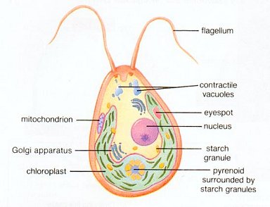
Figure 08a Green Algae
[view large image]

Figure 08b Plant Cell
[view large image]

Subsequent fusion with mitochondria generated cells ancestral to those of animals and fungi. Acquisition by cyanobacteria in yet another round of fusion launched the photosynthetic protists and plants.
It was against this background the green algae emerged as the ancestral to the first plants (see Figure 02). There are single-cells, colonial, filamentous, and multicellular green algae. Algae are grouped according to their pigmentation (green, golden brown, brown, and red) and biochemical differences, such as the chemistry of the cell wall and the chemical compound used to store excess food. Figure 08a shows the structure of a motile green alga - the Chlamydomonas. Figure 08b depicts the structure of a modern plant cell. Comparison shows that all the major organelles in the plant cell are already in place in the green algae. The green algae also have some of the same characteristics as plants. All types of plant life cycles (see Figure 10-06) are seen in this diverse group. For example, the multicellular Ulva (sea lettuce) shows the alternation of generations life cycle in the same way as terrestrial plants. Since the diplontic life cycle involves the fusion of two gametes, the green algae must have learnt about sex already. Actually sexual reproduction took a long time to evolve, it started about 1.2 billion years ago and there have been many intermediate stages in its working out. Protists and some plants to this day maintain the alternative binary fission and sexual reproduction. Incidentally, sex is not equal to reproduction - there can be sex without reproduction such as in abortion or there can be reproduction without sex such as cloning. The two processes are connected only in sexual reproduction.

Symbiotic relationship between organisms is much alive today. A modern example of bacteria incorporating into vertebrate cells is provided by the 2010 study of spotted salamander embryo containing algae. The bright green color from the embryos themselves, as well as from the enclosing capsule betrays the presence of the algae as shown in Figure 09. It seems that the nitrogen-rich waste produced by the embryo is useful to the algae, and the algae in turn supply the oxygen for the respiring embryos. Such a close co-existence with a photosynthetic organism has previously been found in invertebrates, but never in a vertebrate; because vertebrate cells have the adaptive
Salamander Embryos immune system, which destroys biological material not considered "self". But in this case, the salamander cells have either turned off their internal immune system, or the algae have somehow bypassed it. Experiment reveals that the algae gain entry into the embryo when its nervous systems begin to form. The presence of algae in the oviducts of adult female spotted salamanders raises the possibility that symbiotic algae are passed from mother to the offspring's jelly sacs during reproduction. Another intriguing possibility is for the genome of the alga incorporated into the salamander germ cells.

Figure 09 Salamander Embryos Co-existing with Algae [view large image]

Biosphere is defined as a closed system including all living thing (see Figure 10). However, the components within do not live in isolation. They interact with each other in the process called symbiotic relation which can be mutually beneficial to both sides, only one side or neutral.
Symbiotic In case of extremely mutual benefit, it becomes endo (within/inside) - symbiotic which is achieved in a very complicated process including passing the trait to later generations. The 2024 experiment "Bacteria implanted in fungi hints at ancient relationships that helped cells evolve" tried to animate the process with only minimal success even by lot of manipulations.

Figure 10 Symbiotic [view large image]

The "Human Microbiome" is a living example of symbiotic in all of us.
See "Clownfish" for mutualism.
Here are some examples of endo-symbiotic relationships by courtesy of ChatGPT (in Italic) :

1. Mitochondria and Eukaryotic Cells
" Description: Mitochondria, the powerhouse of eukaryotic cells, are thought to have originated from an ancient endosymbiosis event between a primitive eukaryotic cell and an alpha-proteobacterium.
" Benefit: The host cell gained the ability to efficiently produce ATP through oxidative phosphorylation, while the bacterium received protection and a stable environment.
" Key Feature: Mitochondria have their own genome, which is highly reduced and encodes proteins essential for mitochondrial function, reflecting their bacterial ancestry.
2. Chloroplasts and Plant Cells
" Description: Chloroplasts, found in plants and algae, originated from a symbiotic relationship between a eukaryotic cell and a photosynthetic cyanobacterium.
" Benefit: The host cell gained the ability to perform photosynthesis and produce energy from sunlight, while the cyanobacterium obtained a secure environment and access to resources.
" Key Feature: Like mitochondria, chloroplasts have their own DNA, which is also highly reduced compared to their free-living cyanobacterial relatives.
3. Wolbachia and Insects/Nematodes
" Description: Wolbachia is a genus of bacteria that infects many species of insects and some nematodes, living within their cells.
" Benefit: The relationship can range from parasitic to mutualistic, depending on the host species. Wolbachia can influence host reproduction and, in some cases, confer benefits like increased resistance to viruses.
" Key Feature: Wolbachia can manipulate host reproductive mechanisms, such as causing cytoplasmic incompatibility, which increases its transmission through the host population.
4. Aphids and Buchnera Bacteria
" Description: Aphids have an endosymbiotic relationship with the bacterium Buchnera aphidicola, which lives in specialized cells called bacteriocytes.
" Benefit: Buchnera provides essential amino acids that aphids cannot obtain from their sap-based diet, while the aphids provide a protected environment and nutrients to the bacteria.
" Key Feature: Buchnera has a highly reduced genome, losing many genes that are no longer necessary due to its stable environment within the host.
5. Riftia Tubeworms and Chemosynthetic Bacteria
" Description: The giant tube worm Riftia pachyptila lives near hydrothermal vents and houses chemosynthetic bacteria within its trophosome organ.
" Benefit: The bacteria convert toxic hydrogen sulfide into organic molecules, providing nourishment for the tubeworm, which lacks a digestive system and relies entirely on the bacteria for food.
" Key Feature: This relationship is crucial for survival in the extreme, nutrient-poor environment of hydrothermal vents.
6. Termites and Gut Microbes
" Description: Termites have a complex community of protists and bacteria living in their hindguts that help them digest cellulose from wood.
" Benefit: The microbes break down cellulose into simpler compounds that termites can absorb, providing them with essential nutrients. In return, the microbes receive a constant supply of cellulose and a stable habitat.
" Key Feature: Some termite gut microbes have developed intricate metabolic interactions, passing metabolic byproducts between different species to achieve efficient cellulose digestion.
7. Coral and Zooxanthellae
" Description: Reef-building corals contain photosynthetic dinoflagellates called zooxanthellae (genus Symbiodinium) within their cells.
" Benefit: The zooxanthellae provide the coral with organic molecules produced through photosynthesis, while the coral provides the symbiont with carbon dioxide and a protected environment.
" Key Feature: The health of this endosymbiotic relationship is crucial for the survival of coral reefs. Coral bleaching occurs when this relationship breaks down under stressful conditions, such as elevated temperatures.
8. Pea Plants and Rhizobia Bacteria
" Description: Leguminous plants (e.g., peas and beans) form root nodules to house nitrogen-fixing bacteria called rhizobia.
" Benefit: The rhizobia convert atmospheric nitrogen into ammonia, which the plant can use for growth. In return, the plant supplies the bacteria with carbohydrates and a niche for colonization.
" Key Feature: This mutualistic relationship is critical for soil fertility and agricultural productivity, as it naturally enriches the soil with nitrogen.

These examples illustrate the diversity of endosymbiotic relationships and how they can evolve from initial interactions that eventually become essential for the survival and function of both partners.

Go to Next Section
 or to Top of Page to Select
 or to Main Menu

.