

| Home Page | Overview | Site Map | Index | Appendix | Illustration | About | Contact | Update | FAQ |
 |
 |
reinforced in modern time by such similarity. While unicelluar organisms can acquire nutrients and expel waste directly in aquatic environment, they could not survive on dry surfaces with a humidity of less than 10%. The same situation is applicable to the cells in multicelluar organisms which solve the problem with the "Internal Sea" in the form of Interstitial Fluid (ISF). |
Figure 01 Origin of Life |
Figure 02 Sea Water and Body Fluids, |
Figure 02 also shows that the fluid composition inside the cell (Intracellular Fluid, ICF) is quite different from sea water as the constituents are altered by the metabolic process of life. |
 |
40% in the elderly (Figure 03). There are two kinds of body fluids, namely, the intracellular (ICF) and extracellular (ECF) which is further categorized into plasma and interstitial fluid (ISF). Since the source of ISF is derived from the plasma, they have essentially the same composition with lower concentration of proteins in ISF (Figure 02). In addition, small amount of lymph in the lymphatic system and sometimes TSF (Transcellular Fluids such as cerebrospinal, ocular and joint fluids ) are also included into the ECF in some accounting. The composition of ICF varies depending on the type of organ. As illustrated in Figure 03, the lungs contains 90% water while the bones are very dry at 22%. |
Figure 03 Body Fluids |
 |
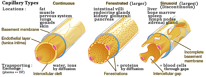
proteins called interstitial matrix. It acts as a compression buffer against the stress placed on the ECM as shown in Figure 04 (right). The interstitial fluid (ISF) is another component that fills the rest of the ECM space providing a vital service for the maintenance of life. Nutrient and waste from and to the ISF must diffuse across the basement membrane or through gaps (for the discontinuous type, see Figure 05) to be absorbed by the cells. |
Figure 04 Connective Tissue and (ECM) Extracellular Matrix [view large image] |
epi-: prefix taken from the Greek that means "on, upon, at, by, near, over, on top of, toward, against, among." |
 |
 |
The exchange is facilitated through the difference between the hydrostatic and osmotic pressures (see Figure 06, top). About 2% of the ISF is drained via the lymphatic system, which has no pumping "heart". It depends on body movements to force the fluid into the lymphatic capillaries. All the cells in multicellular organisms obtain nutrient and remove waste through the ISF - the "Internal Sea" (serum in medical term). |
Figure 05 Types of Capillaries [view large image] |
Figure 06 Global View of ECF [view large image] |
BTW, red blood cells and platelets cannot pass through the walls of the capillaries (too large). |
 |
|
Figure 07 Interstitial Fluid (ISF) [view large image] |
|
 |
The cells in various organs are at the other end of ISF running the reverse process (for capillary) of receiving nutrient and rejecting waste. There are many methods according to the kind of substance. Transport of smaller molecules depends on concentration gradient as shown in Figure 08a. Figure 08b provides a global view of the salt and water transport through the cellular membrane. The larger molecules are transported via a more elaborate process (Figure 09). |
Figure 08 Cell Transport [view large image] |
The three types of cellular transport for smaller molecules are explained briefly below. |
 |
|
Figure 09 Transcytosis |
 |
Edema is an abnormal accumulation of fluid in ISF manifesting as swelling. The amount of ISF is determined by the balance of fluid homeostasis, which controls the proportion of plasma and ISF in the ECF to a range of (20% - 25%) and (80% - 75%) respectively. The increased secretion of fluid into ISF, or the impaired removal of the fluid, can cause the condition. The symptoms vary according to the location where the swelling occur (see article by "Mayo Clinic"). |
Figure 10 Oedema [view large image] |
Figure 10 shows a case of leg swelling caused by blocking or loss of the lymphatic capillary. |
 |
 |
While the effect of mechanical force on embryonic development has been demonstrated conclusively by many experiments. The most common examples of hydrostatic pressure transmission are the various kinds of massage, which facilitate the circulation of interstitial fluid by mechanical force. Meditation helps to promote recycling of the fluid by relaxing the muscles as well as the mind. Qi-gang directs the pressure to move along a direction (need supervision to prevent blockage or bleeding through wrong pathway). Then some forms of martial art use the internal fluid pressure against external force (Figure 12). |
Figure 11 |
Figure 12 Hydrostatic Pressure Transmission, Effect of |
See also ""Mechano-transduction". |
 |
Examples include oxygen, carbon dioxide, and lipid-soluble molecules like certain vitamins. On the waste side, simple diffusion allows the movement of small waste molecules, such as water and urea, out of the cell if there's a higher concentration inside the cell. Composition of the cell membrane plays a crucial role in regulating what can and cannot pass through. Thus, charged or polar substances cannot go through, because most of the lipids in the cell membrane are phospholipids. Each phospholipid molecule has a polar head and 2 nonpolar tails. When surrounded by water, phospholipid molecules form a bilayer naturally. The heads, being polar, are attracted to the water (hydrophillic), which is also polar; therefore, the heads face outward. The nonpolar tails face inside, away from the water (hydrophobic). Such kind of struction prevents polar molecules to go through. |
Figure 13 Transport Processes [view large image] |
 |
|
Figure 14 Proton Pump |
 ADP + Pi + 0.32 ev ----- (7).
 |
|
Figure 15 Sodium Pump in Action [view large image] |
Figure 15 also suggests a possible reverse direction to produce ATP. It involves only the backward direction from (3) to (2), the shape of the pump and concentrations remain unchanged. The process promotes the ADP ground state to the ATP meta-stable state (see insert in lower left corner). In effect, the pump just serves as an enzyme to speed up the backward reaction. |
 |
Ultimately, the energy stored in the electrochemical gradient is created by the ATP synthase. It's like hitching a ride on the energy coattails of another process. There are two different kinds of gradient : * The chemical gradient, or difference in solute concentration across a membrane. * The electrical gradient, or difference in charge across a membrane. As shown in Figure 16, antiport moves 2 different species in opposite directions, while symport let them go in same direction. Uniport simply allows only one species across from low to high concentration. |
Figure 16 Secondary Active Transport [view large image] |
 |
 |
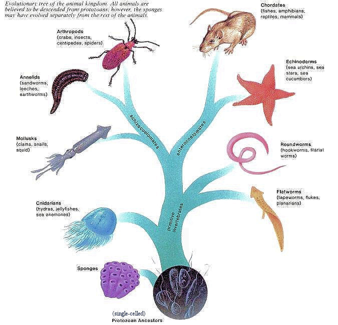
All single-celled protozoa use the above mentioned mechanism (the various kinds of transport) to acquire nutrition and expel waste. Each cell in mulit-cellular animals (see main branch in Figure 17) also runs in this way. However, those cells are no longer in a "sea water" environment, their life now depends on the Interstitial Fluid (ISF) which is supplemented by some sort of circulatory system (via heart, blood, and lymph, see Figure 18 for an advanced system) and getting more sophisticated in more advanced phyla as the animals evolved. The following table summarizes how it is done by different phyla starting from the very primitive sponges with none of those apparatuses. |
Figure 17 Animal Evolution |
Figure 18 |
| Animal | Circulatory System | Heart | Blood | Lymph | |
|---|---|---|---|---|---|
| Sponges | The amoeboid cells (having changeable shape) within the wall act as a circulatory device to transport nutrients and wastes from cell to cell. This is something liike "personal service", no need for ISF. | No | No | No | |
| Cnidarians | Nutrients and wastes in ISF are passed by diffusion to the rest of the body from gastrodermis cells. | No | No | No | |
| Flatworms | Since the body is flattened, nutrients are easily passed by diffusion from cell to cell via ISF. | No | No | No | |
| Roundworms | Nutrients and waste are distributed in the body cavity as ISF, whose contents are regulated by one-celled glands or excretory canal along each side of the body. | No | No | No | |
| Mollusks - Clam/Snail | They have heart that pumps blue blood (containing pigment hemocyanin instead of red hemoglobin) which leaks from the vessels into sinuses once in the organs. | Yes | Blue blood into sinuses | No | |
| Mollusks - Octopus | Octopuses have blue blood and 3 hearts, 2 at the end of each gills; they pump blood through the gills. The third heart pumps blood through the rest of the body. | 3 hearts | Blue blood | No | |
| Annelids | The earthworm has an extensive closed circulatory system. Hemoglobin-containing blood moves anteriorly in a dorsal blood vessel and then is pumped by five pairs of hearts into a ventral blood vessel. As the ventral blood vessel takes blood toward the posterior regions of the worm's body, it gives off branches in every segment. | 5 pairs of hearts | Yes | No | |
| Arthropods | They have an open circulatory system. Colorless blood is pumped dorsally from the heart and then enters sinuses and hemocoel, where it comes in direct contact with the tissues (see cockroach) | Yes | Colorless blood | Yes | |
| Echinoderms See gene-expression. 2023 |
The echinoderms have an open circulatory system. They have no heart. Coelomic fluid, circulated by ciliary action, performs many of the normal functions of a circulatory system. The system consists of a central ring canal and radial canals extending along each arm. Water (via the tube feet) circulates through these structures allowing for gas, nutrient, and waste exchange. | ~ Cardiac stomach | ~ Coelomic fluid | No | |
| Fishes | The heart of a fish is a simple pump, and the blood flows through the chambers, including a non-divided atrium and ventricle, to the gills only. Oxygenated blood leaves the gills and goes to the body proper. | Yes, 2 chambers |
Yes, cold blooded |
Some; see "Fish SVS" |
|
| Amphibians | Their circulatory system now changes to refresh the blood by lung with a 3 chambers heart, which is not very efficient by mixing purified and dirty blood there. So the blood receives further oxygenation by Cutaneous Gas Exchange via the skin. | Yes, 3 chambers |
Yes, cold blooded |
Yes | |
| Reptiles | Construction of the 3 chambers is different from the amphibians' with the 3rd isolated from the lung. It is for diving in aquatic species, e.g., in sea turtles and sea snakes, bypassing the lungs could prevent gas entering the blood stream to avoid sickness. | Yes, 3 chambers |
Yes, cold blooded |
Yes, Lymph heart |
|
| Birds | The avian heart resembles the Mammalian (human) heart, with four chambers and valves. However, the inner walls of the atria and ventricles are smoother in birds than in humans, and the avian valves are simpler than their mammalian counterparts. | Yes, 4 chambers |
Yes, warm blooded |
Yes, Lymph heart - flightless birds |
|
| Mammals | The 4 chambers illustration shows that the left half of the mammalian heart differs considerably from the right half. It is found that the mammalian heart is evolved from the primaeval fish's by difference in gene expressions showing the unity in life. | Yes, 4 chambers |
Yes, warm blooded |
Yes, see lymphatic System |

 |
|
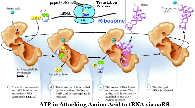
Figure 19 ATP in Action [view large image] |
 |
|
Figure 20 Eco-cycle [view large image] |
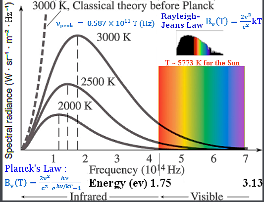
 D2, it is called intensity I = L/4
 D2 = 1.36x10⁶ erg/sec-cm2 = 0.85x10¹⁸ ev/sec-cm2 for the power received every square-cm on Earth from the Sun - aka Solar Constant.
 |
 |
This property of carbon, and the fact that carbon nearly always forms four bonds to other atoms, accounts for the large number of known compounds including glucose. At least 80 percent of the 5 million chemical compounds registered as of the early 1980s contain carbon. The affinity of carbon for the most diverse elements does not differ very greatly - so that even the most diverse derivatives need not vary very much in energy content. This ability allows the organic world to exist in a special form of thermodynamic stability. Figure 22,d shows the location of this very special energy in the solar spectrum. |
Figure 21 Energy from the Sun |
Figure 22 SP3 State of Carbon Atom |
 |
The stand alone SP3 excited state has a half-life about 10-15 sec, the tetrahedra would break down and return to equilibrium in absence of energy infusion in the form of Sun light to have it recycled (see "Photosynthesis - Respiration"). The stability of the SP3 state in organic molecules like carbohydrates (glucose) is maintained by the strength of the covalent bonds and is not subject to spontaneous de-excitation for the individual carbon atom (Figure 23a). The energy stored in SP3 is released with the break up of the covalent bonds. |
Figure 23a SP3 State in Glucose |
The 2 ev energy released by de-excitation of SP3 is distributed in 6 ATP ~ 0.32 ev each.
ChatGPT provides further details (in italic) : |
 |
 |
|
Figure 23 Photosynthesis |
Figure 24 CO2 in Europa |
 |
 |
Legend : RuBP = ribulose-1,5-bisphosphate; RuBisCO = Ribulose-1,5-bisphosphate carboxylase/oxygenase (carbon fixation); 2P-glycolate = 2-phosphoglycolate (oxygen consumption); 3PG = 3-phosphoglycerate; G3P = glyceraldehyde-3-phosphate. |
Figure 25 CO2 over the Eons [view large image] |
Figure 26 Photosynthesis and Calvin Cycle [view large image] |
The (ADP+Pi)/NADP+ - ATP/NADPH cycle is to refresh (ADP+Pi) back to ATP after it is used (as 1 Pi molecule is detached in the process yielding ~ 0.32 ev). |
 |
 |
Figure 28 shows the anoxygenic process to be somewhat similar to the oxygenic photosynthesis completed with the 1st and 2nd subpathways. It also requires Sun light (P840) to run. Here's a comparison for both types : Oxygenic : CO2 + H2O + Energy  O2 + CH2O O2 + CH2ONeglecting the fine details, this process just removes the C from CO2 and appends it to H2O, + O2 Anoxygenic : CO2 + 2H2S + Energy  2S + H2O + CH2O 2S + H2O + CH2ONeglecting the fine details again, this one release the 2S, appends 1 H2 to 1 O (in CO2) to form H2O; CH2O is generated by joining the remaining CO to the other H2. |
Figure 27 Pre-Cambrian Era [view large image] |
Figure 28 Anoxygenic Photosynthesis |
BTW, the O atom is similar to the S atom which has just an additional electron shell (see "Periodic Table"). |
 |
 |
Fermentation allows glucose to be continuously broken down to make ATP due to the recycling of NADH to NAD+. Bacteria (yeast etc.) perform fermentation, converting carbohydrates into ethanol (alcohol) according to the formula : C6H12O6 (glucose) ------>2 C2H5OH (ethanol) + 2 CO2 See Figure 30,b for more detail. |
Figure 29 Fermentation types [view large image] |
Figure 30 Fermentation [view large image] |
There is another form producing useless lactic acid that leads to muscle fatigue and soreness after strenuous exercise; however, such backup (respiration) process allows continuation of the activity (at slower pace) when the oxygen level is running low in the body (see Figures 29, 30). |
 |
1. Increased chlorophyll content: To capture as much available light as possible, underwater plants may have higher concentrations of chlorophyll in their photosynthetic tissues. 2. Thin and flexible leaves: Thinner and more flexible leaves can allow for better light penetration, enabling the plant to capture sunlight even in low-light conditions. 3. Red pigments: Some underwater plants have red pigments that help them absorb blue and green light, which can penetrate deeper into water than other colors. 4. Vertical orientation: Plants may orient their leaves vertically to optimize light capture, especially in environments where light is limited. 5. Floating leaves: Some water-dwelling plants have floating leaves that position themselves at the water's surface, maximizing exposure to sunlight. Different plants have varying adaptations to low-light conditions and can thrive at different depths. Some aquatic plants can survive and photosynthesize at relatively deep depths. For example, certain types of seagrasses and algae can grow at depths of several meters in clear water. Beyond a certain depth, the intensity and quality of light may not be sufficient to support photosynthesis. In such cases, aquatic plants may not be able to survive, and other forms of life adapted to low-light or dark conditions, such as certain bacteria and fungi, may dominate. |
Figure 31 Water in Plant [view large image] |
 |
1. (O2).- : Superoxide anion 2. H2O2 : Hydrogen peroxide 3. OH.- : Hydroxyl radical 4. NOX : NAD(P)H oxidase The defences : |
Figure 32 Oxygen [view large image] |
1. SOD : SuperOxide_DismutaseSOD 2. Catalase to neutralize H2O2 |
 C6H12O6 + 6O2
 C12H22O11 + H2O.
 |
Our daily diet of carbohydrates are the sugars, starches and fibers found in fruits, grains, vegetables and milk products (Figure 33). They provide fuel for the central nervous system and energy for working muscles, i.e., they are used for respiration (the reverse of photosynthesis). Carbohydrates are digested in the mouth, stomach and small intestine. The amylase and other enzymes break down starch into sugars for assimilation by the cell. |
Figure 33 Carbohydrate |
 |
Overall, carbohydrates play a crucial role in the function and survival of living organisms. |
Figure 34 Digestion |
 |
As shown in a typical eukaryotic cell (Figure 35,c), mitochondria is an organelle in eukaryotic cells - a power house to supply energy for maintaining life in every one of these cells. The macro-view of the process is shown in Figure 35,a with the detailed mechanism shown in Figure 35,b (see glycolysis and "proton pump" for further explanation). Mitochondria has its own genome in the form of a ring similar to those in bacteria. In fact, the functioning of their live machinery as well as appearance are similar to those of bacteria. Such kinds of observations has prompted the theory of extracellular origin for mitochondria in the 1970s. This view is now widely accepted by biologists today. |
Figure 35 Mitochondria in Respiration [view large image] |
Actually, the bacteria do not have all the organelles in the eukaryotic cell (see "PKC in Table 11-02") |
 |
Among the multitude of micro-organisms, two bacteria destined to become the major components for the further development of the living world. The mitochondria (see Figure 36) are the sites of the principal oxidation reaction linked to the assembly of ATP, which supplies energy to most of the organisms today. Their closest relative among present-day bacteria have been identified to be the alpha-proteobacteria. The chloroplasts (see Figure 26) are the agents of photosynthesis in unicellular algae and plants. They store the solar energy in the form of glucose (sugar), which becomes food for the "heterotrophs". Their present-day relative are the cyanobacteria (formerly known as blue-green algae), which is believed to be responsible for the first generation of atmospheric oxygen. As shown in Figure 33 the chloroplasts supply food and oxygen for the heterotropic organism, which in turn produced CO2 for photosynthesis. This ecocycle generates a lot of atmospheric oxygen as shown in Figure 32,c today. The evolutionary steps for the micro-organisms are shown in Figure 27, which identifies the first organisms to be anaerobic (anoxygenic) autotrophy. |
Figure 36 Mitochondria [view large image] |
Actually, the enlarged view of the mitochondria in Figure 36 reveals that it doesn't look like a cell anymore. Only a few organells (not shown) and a plasmid DNA remain. For example, it has ribosomes and accessories such as tDNA, mDNA etc. to produce proteins as enzymes in ATP production. |
 |
Each mitochondria has numerous copies of circular, double-strand plasmid DNA cosising of a 16,569 nucleotide sequence. The sequence contains a total of 37 genes for making proteins, tRNA, and rRNA (see Figure 37). The 13 protein-coding genes are not enough for producing all the mitochondria-specific proteins. It is surmised that the billion years' symbiotic relationship with the hosts progressively reduced its independence by transferring many of its genes to the nuclear genome (of the host). Thus, many mitochondrial disorders are related to mutations in the nuclear genes - a price to be paid by un-loading burdens to the "central government" (so to speak). Actually, the host is happy to oblige so that the mitochondria can concentrate on making the all important ATP to meet the energy requirement. |
Figure 37 Mitochondrial DNA [view large image] |