Home Page Overview Site Map Index Appendix Illustration About Contact Update FAQ


Mulitcellular Organisms


Hematopoiesis (Formation of Blood Cells), and Lymphatic System

Contents

Bones
Bone Marrow and Blood Types
Hematopoiesis Progression
Variation in Species
Lymphatic System

Bones

Bones 
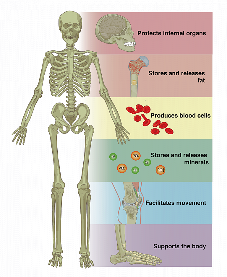
Bones can persist for a long time, potentially thousands of years, under favorable conditions like dry environments or in certain types of soil. Generally, there are 4 different types (see Figure 10-13h1); the long bone is the strongest. Bones serve many vital function (see Figure 10-13h2); here's a list provided by ChatGPT (in italic) :
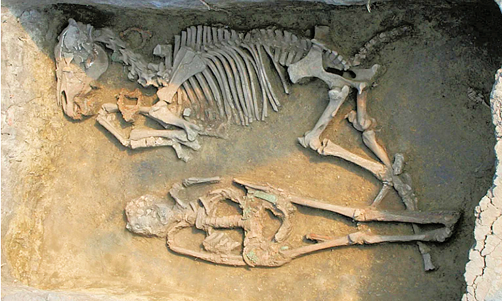
Bone Fossil

1. Support: Bones provide structural support for the body, allowing vertebrates to maintain their shape and posture. The skeleton acts as a framework to support soft tissues and organs.
2. Protection: Bones protect vital organs and tissues from injury. For example, the skull protects the brain, the ribcage protects the heart and lungs, and the vertebrae protect the spinal cord.
3. Movement: Bones, along with muscles and joints, facilitate movement. Muscles attach to bones via tendons, and when muscles contract, they pull on the bones, causing movement.
4. Mineral Storage: Bones store minerals such as calcium and phosphorus, which are essential for various physiological processes in the body. During times of need, such as when calcium levels are low, bones release minerals into the bloodstream to maintain homeostasis.
5. Energy Storage: In addition to minerals, bones also store energy in the form of lipids (fats). Yellow marrow, found in the central cavities of long bones, consists mainly of adipocytes (fat cells) and serves as a reservoir of energy.
Bones Bone Functions 
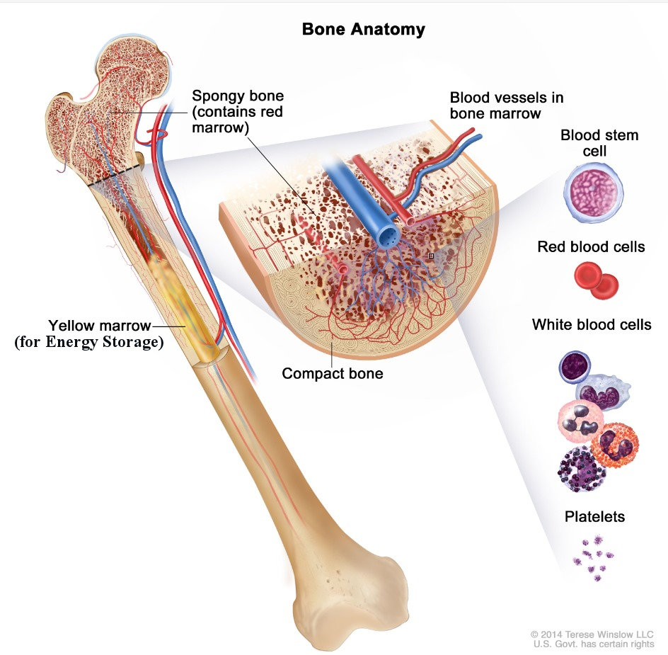
6. Blood Cell Production: Within the bone marrow, specialized cells called hematopoietic stem cells produce various types of blood cells, including red blood cells, white blood cells, and platelets. This process is known as hematopoiesis and occurs primarily in the marrow of certain bones, such as the pelvis, sternum, and long bones. The red marrow found within these bones contains hematopoietic stem cells that continuously produce red blood cells, white blood cells, and platelets throughout adulthood.

Figure 10-13h1 Types of Bone

Figure 10-13h2 Uses

As for the evolution of skeleton, the "exoskeleton to endoskeleton hypothesis" asserts that the bony structures (including teeth) were evolved from hard shell, scales or armor outside turning inward inside the body (Figure 10-13h3).

This idea has been proposed to explain the evolutionary transition from early vertebrates with an external bony armor to more advanced vertebrates with an internal skeletal system.there are several lines of evidence that support aspects of this hypothesis:

1. Developmental similarities: During embryonic development, there are similarities in the formation of both external and internal skeletal structures, suggesting a common evolutionary origin.
2. Genetic evidence: Studies of the genetic regulation of skeletal development have identified shared genetic pathways between the development of external and internal skeletal structures, supporting the idea of a common evolutionary origin.
3. Fossil evidence: Fossil discoveries have revealed transitional forms between ancient fish-like creatures with external armor and early vertebrates with internal skeletons, providing some support for the idea of a gradual transition from exoskeleton to endoskeleton.

Skeleton This transition may have provided several advantages, such as increased flexibility, greater mobility, and improved protection of internal organs. The shift from exoskeleton to endoskeleton likely occurred gradually over evolutionary time, driven by environmental pressures and the need to adapt to changing habitats and ecological niches.

Figure 10-13h3 Skeleton

See a review article on "Where did bone come from?"

[Top]


Bone Marrow

During adulthood, the primary sites of hematopoiesis in humans are the bones of the pelvis, sternum, ribs, skull, and the proximal ends of the long bones such as the femur and humerus. Figure 10-13h4 shows the structure of a long bone (the femur). The blood cell production site is in the red marrow which makes differenr kinds of cell as shown in Figure 10-13h5 and listed by ChatGPT (in italic) :

1. Red Blood Cells (Erythrocytes):
* Function: Transport oxygen from the lungs to tissues throughout the body and carry carbon dioxide back to the lungs for exhalation.
* Structure: Red blood cells are small, biconcave discs without a nucleus. Their shape allows for efficient gas exchange.
* Lifespan: Red blood cells typically live for about 120 days before being removed by the spleen and liver.
* Types :
1. Presence of Antigens (A and B):
+ Type A blood has A antigens on the surface of its red blood cells.
+ Type B blood has B antigens on the surface of its red blood cells.
+ Type AB blood has both A and B antigens on the surface of its red blood cells.
+ Type O blood has neither A nor B antigens on the surface of its red blood cells.
2. Presence of Antibodies:
+ Type A blood has anti-B antibodies in the plasma.
+ Type B blood has anti-A antibodies in the plasma.
+ Type AB blood has neither anti-A nor anti-B antibodies in the plasma.
+ Type O blood has both anti-A and anti-B antibodies in the plasma.
3. Compatibility:
+ Type A blood can donate to individuals with type A or AB blood and can receive from individuals with type A or O blood.
+ Type B blood can donate to individuals with type B or AB blood and can receive from individuals with type B or O blood.
+ Type AB blood can donate to individuals with type AB blood and can receive from individuals with any blood type (universal recipient).
+ Type O blood can donate to individuals with any blood type (universal donor) but can only receive from individuals with type O blood.

These differences are crucial in blood transfusions to ensure compatibility and prevent adverse reactions, such as hemolysis (the destruction of red blood cells). Additionally, there are other blood group systems, such as the Rh system (Rh factor), which further refine blood typing and compatibility.

2. White Blood Cells (Leukocytes):
* Function: White blood cells are a crucial part of the immune system, defending the body against infections and foreign invaders.
* Types:
+ Neutrophils: Phagocytic cells that engulf and destroy bacteria and other foreign particles.
+ Lymphocytes:
   B cells - Produce antibodies to neutralize pathogens and mark them for destruction.
   T cells - Directly attack infected or abnormal cells and regulate the immune response.
+ Monocytes: Precursors to macrophages, they engulf and digest pathogens, dead cells, and debris.
+ Eosinophils: Primarily involved in combating parasitic infections and modulating allergic responses.
+ Basophils: Release histamine and other inflammatory mediators in response to allergic reactions and parasitic infections.

Bone Marrow Blood Cells 
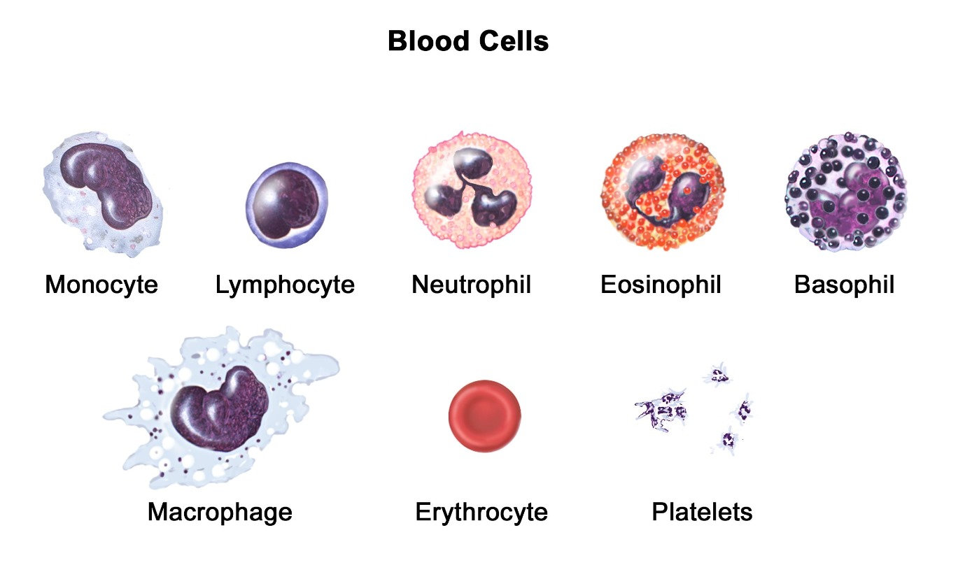
3. Platelets (Thrombocytes):
* Function: Essential for blood clotting (hemostasis) to stop bleeding when blood vessels are damaged.
* Structure: Platelets are small, irregularly shaped cell fragments derived from the fragmentation of megakaryocytes in the bone marrow.
* Role in Hemostasis: Platelets adhere to the site of vascular injury, aggregate to form a plug, and release factors that promote blood clot formation.

Figure 10-13h4 Bone Marrow

Figure 10-13h5 Blood Cells [view large image]

These blood cells work together in a coordinated manner to maintain the body's health and protect against pathogens and injuries. It becomes fatal if blood loss is over 40%.
See "Powerful imaging shows blood cells made in bone" and Figure 10-13h6
Blood Cell Production The March, 2024 article "Resilient anatomy and local plasticity of naive and stress haematopoiesis" on haematopoiesis response to "insult" (such haemorrhage, bacterial infection and during ageing) is followed by the April, 2024 article "Depleting myeloid-biased haematopoietic stem cells rejuvenates aged immunity", which demonstrates that increasing certain type of Haematopoietic Stem Cell (HSC) would enable aged mice to restore characteristic features of a more youthful immune system. It seems that "haematopoietic" is an active research subject because it is directly related to aging (see Figure 10-13h7).

Figure 10-13h6 Production

[Top]


Hematopoiesis Progression

Bone Marrow The production sites of hematopoiesis, where blood cells are formed, changes throughout a person's lifetime due to developmental and physiological factors. The progression involves the commplicated interaction between 2 types of stem cells, see "Hematopoiesis during Ontogenesis, Adult Life, and Aging". Figure 10-13h7 shows the changes during the lifetime in term of cellularity (number of cells in a given tissue sample in term of precentage), while ChartGPT provides a short description (in italic) :

Figure 10-13h7 Progression

1. Embryonic Development: During early embryonic development, hematopoiesis primarily occurs in the yolk sac, where the first blood cells are formed. As development progresses, hematopoiesis shifts to other sites.
2. Fetal Development: In the mid-gestation period, hematopoiesis transitions to the fetal liver, which becomes the primary site for generating blood cells. The fetal liver has a rich blood supply and provides the necessary environment for hematopoietic stem cells to proliferate and differentiate.
3. Birth and Infancy: After birth, hematopoiesis occurs mainly in the bone marrow, particularly in the long bones such as the femur and tibia. The bone marrow contains specialized niches that support the proliferation and differentiation of hematopoietic stem cells into various blood cell types.
4. Adulthood: Throughout adulthood, the bone marrow remains the primary site of hematopoiesis. However, as the body ages, there can be changes in the composition and function of the bone marrow microenvironment, which may affect hematopoietic activity. Additionally, in certain circumstances, extramedullary hematopoiesis (hematopoiesis outside the bone marrow) can occur, such as in the spleen or liver, particularly in response to certain diseases or conditions.
5. Old Age: In older adults, there can be alterations in the bone marrow microenvironment, including a decline in hematopoietic stem cell function and changes in the composition of supportive stromal cells. This can result in decreased hematopoietic activity and may contribute to age-related changes in the immune system, such as immunosenescence.

Overall, the varying sites of hematopoiesis reflect the dynamic nature of blood cell production throughout different stages of development and aging, as well as the adaptability of the hematopoietic system to changing physiological demands.

[Top]


Variation in Species

According to ChatGPT (in italic) :

The process of blood cell formation, occurs in various organs across different species of vertebrates. Here's a list of some of these organs in different vertebrate groups:

1. Mammals:
* Bone Marrow: In mammals, including humans, the primary site of hematopoiesis is the bone marrow. It can be found in both flat bones (such as the sternum and pelvis) and long bones (such as the femur and humerus).
* Fetal Liver and Spleen: During fetal development, hematopoiesis can also occur in the liver and spleen.
2. Birds:
* Bone Marrow: Similar to mammals, birds have hematopoietic activity in their bone marrow.
* Spleen: The spleen also plays a role in hematopoiesis in birds.
3. Fish:
* Kidney: In many fish species, the kidney is the primary site of hematopoiesis.
* Spleen: The spleen in fish also contributes to hematopoiesis.
* Head Kidney: Some fish species have a specialized hematopoietic tissue called the head kidney, which is involved in blood cell production.
4. Amphibians:
* Liver: In amphibians such as frogs, the liver is one of the major sites of hematopoiesis.
* Spleen: Similar to other vertebrates, the spleen also plays a role in amphibian hematopoiesis.
5. Reptiles:
* Bone Marrow: Hematopoiesis occurs in the bone marrow of reptiles.
* Spleen: The spleen may also contribute to hematopoiesis in reptiles.

These are general patterns, and there can be variations among species within each group. Additionally, in some cases, hematopoietic activity may change during different life stages or in response to environmental factors.


It shows that the hematopoiesis site is located in the spleen (see Figure 10-13h8) for all species of vertebrates except the mammal. Even in human, the spleen manages all the functions about blood (except hematopoiesis in adult) including :

* Storing blood.
* Filtering blood by removing cellular waste and getting rid of old or damaged blood cells.
* Making white blood cells and antibodies to fight infection.
* Maintaining the levels of fluid in the body.
* Producing antibodies for protection against infection.

Spleen The preference for bone marrow over the spleen in mammals may be due to factors such as the bone marrow's larger capacity for hematopoiesis and its more specialized microenvironment, which supports the proliferation and differentiation of hematopoietic stem cells. Additionally, the bone marrow provides a more protected environment for blood cell formation compared to the spleen, which is more susceptible to damage and infection.
Actually, the lymph node in human can be considered as the miniature version of the spleen (see Figure 10-13-h6).

Figure 10-13h8 Spleen

Lymphatic System - Lymph nodes are essential components of the system, serving several crucial functions:

Lymph Nodes 
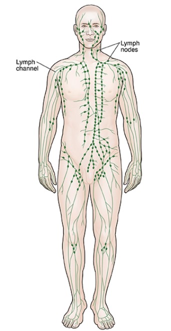
1. Filtration: Lymph nodes act as filters, trapping and removing foreign particles, such as bacteria, viruses, and cellular debris, from the lymph fluid before it returns to the bloodstream. This helps to prevent the spread of infections and diseases throughout the body.
2. Immune response: Lymph nodes are hubs for immune cell activation and coordination. They contain various types of immune cells, including lymphocytes (B cells and T cells), macrophages, and dendritic cells. When foreign substances are detected in the lymph fluid, immune cells within the lymph nodes become activated, initiating an immune response to eliminate the threat.
3. Production of lymphocytes: Lymph nodes support the production and maturation of lymphocytes, which are crucial for the adaptive immune response. B cells mature and differentiate into plasma cells
(which make antibodies to fight bacteria and viruses, to stop infection and disease).

Figure 10-13h9 Nodes

[Top]


Lymph Fluid

Lymph fluid circulates through the lymphatic system primarily due to three main factors:
1. Skeletal Muscle Contractions: The lymphatic vessels rely on the contraction of nearby skeletal muscles to push lymph fluid along. As muscles contract during movement (walking, exercising, etc.), they compress the lymphatic vessels, pushing the lymph fluid forward.
2. Valves in the Lymphatic Vessels: Similar to veins, lymphatic vessels contain one-way valves that prevent the backflow of lymph fluid. As the skeletal muscles push the fluid, the valves ensure it moves in the correct direction toward the larger lymphatic vessels and ultimately drains into the bloodstream.
LymFluid 3. Pressure Changes from Breathing: The act of breathing creates pressure changes in the thoracic cavity (chest). These changes help move lymph fluid, especially through the larger lymphatic ducts (like the thoracic duct), as the pressure gradients assist in drawing the lymph upward toward the subclavian veins, where it reenters the bloodstream.
4. Body Position: Gravity can affect lymph flow, particularly in the lower extremities.

Figure 10-13h10 Lymphatic System [view large image]

See "Lymphatic System- Organs, Functions, Diseases".


Unlike blood circulation, the lymphatic system does not have a central pump like the heart. Instead, these passive mechanisms (muscle contraction, one-way valves, and pressure changes) are essential for moving lymph through the body. In general, the lymphatic system works much more slowly than the cardiovascular system, but its slow pace is sufficient for its role in fluid balance, immune function, and the removal of waste products. The speed of lymph fluid movement is quite slow compared to blood circulation. On average, lymph flows at a rate of about 120 milliliters per hour (0.1 to 0.2 millimeters per second) in the entire lymphatic system. This equates to approximately 0.002 to 0.3 centimeters per second in different regions of the body, depending on factors like proximity to lymph nodes or major lymphatic ducts.

The time it takes for lymph fluid to transport debris to lymph nodes can vary, it generally reaches nearby lymph nodes within a few hours to a couple of days. Here's how it works:
" Local debris: For debris or immune responses near the lymph nodes (e.g., a nearby infection or injury), lymph can transport debris to the nodes within hours to a day.
" Farther areas: If debris is coming from more distant parts of the body, like the lower extremities, it might take a day or two for it to reach major lymph nodes, depending on the flow and activity level of the person.
Circulation 
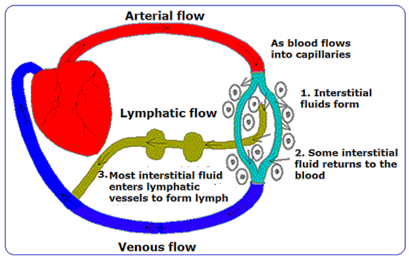
In general, the lymphatic system efficiently cleans up and filters waste within a few days under normal conditions. Physical activity, breathing, and overall health can speed up this process.

Circulation

+ Flow of Lymph Video


Dark moles, also known as melanocytic nevi, are clusters of pigmented cells called melanocytes that form in the skin. The reason they cannot be removed by lymph fluid is due to several factors:

1. Nature of Moles: Moles are made of a dense concentration of melanocytes, which are specialized skin cells that produce melanin, the pigment responsible for skin color. These cells are part of the skin structure and not foreign particles, so the body's immune system doesn't target them for removal like it would for pathogens or damaged cells.
Moles 2. Lymphatic System Function: The lymphatic system primarily helps in removing waste products, toxins, and unwanted materials like bacteria or viruses from the body. It doesn't target healthy, non-pathogenic cells like melanocytes unless there is an abnormal change, such as malignancy.
3. Mole Location: Moles are typically located in the dermis and epidermis layers of the skin, where they integrate with normal skin tissue. The lymphatic system is more involved in the removal of fluids, immune cells, and waste products from deeper tissues rather than surface-level changes like moles.
4. Stable Nature of Most Moles: Most moles are benign, stable structures. Since they don't pose a threat or signal damage, the body has no mechanism to remove them naturally.

Figure 10-13h11 Moles [view large image]

In cases where moles need to be removed, such as for cosmetic reasons or if they show signs of becoming cancerous (e.g., melanoma), they are typically excised surgically.
See more about "Moles".


Older people tend to have more dark moles or pigmented lesions due to several factors related to aging and cumulative sun exposure. Here are the main reasons:
1. Cumulative Sun Exposure: Over time, the skin is exposed to ultraviolet (UV) radiation from the sun. UV radiation can cause changes in melanocytes, the cells responsible for producing melanin (skin pigment). The more sun exposure a person accumulates over their lifetime, the more likely they are to develop dark moles or pigmented lesions. UV damage can cause existing moles to darken or new ones to form.
2. Changes in Skin Cell Turnover: As people age, the skin's ability to regenerate and repair itself slows down. This can lead to an accumulation of pigmented cells, which may cause moles or other pigmented spots to become more prominent or numerous.
3. Hormonal Changes: Hormones can affect melanocyte activity, and hormonal fluctuations over a lifetime can contribute to the appearance of new moles. For instance, hormonal changes during pregnancy, menopause, or even certain medications can trigger the formation of moles.
4. Genetics: Some individuals are genetically predisposed to develop more moles or have them darken with age. This genetic influence may be more apparent as they get older.
5. Aging-Related Skin Changes: Aging leads to overall changes in the structure and function of the skin. The skin becomes thinner, and the mechanisms controlling pigment production can become irregular, leading to a higher likelihood of dark moles or age spots (solar lentigines).

While many dark moles are benign, it's important for older individuals to monitor their skin regularly, as changes in moles can sometimes indicate a risk of skin cancer.

More dark moles do not necessarily indicate a health problem, as many moles are benign. However, certain changes in moles or the appearance of many new ones could be a cause for concern, particularly if there are signs of abnormal growth or potential skin cancer, like melanoma. Here are factors to watch for:
1. Benign Moles
Many dark moles are harmless and result from factors like sun exposure, aging, or genetics. In some individuals, it's normal to have many moles, and their presence alone does not indicate poor health.
2. Signs of Melanoma
If a mole starts changing in shape, size, color, or texture, or if new dark moles appear rapidly, it could be a sign of a problem, especially melanoma, a type of skin cancer. A good way to assess moles is by using the ABCDE criteria:
" A (Asymmetry): One half of the mole does not match the other.
" B (Border): Irregular, scalloped, or poorly defined borders.
" C (Color): Varied colors within the mole, such as shades of brown, black, red, or white.
" D (Diameter): Moles larger than 6mm (about the size of a pencil eraser) may be concerning, though melanoma can be smaller.
" E (Evolving): Any change in the mole over time, including growth, color change, or symptoms like itching or bleeding.
3. Sudden Increase in Moles
An unexpected increase in moles or a sudden appearance of many new dark moles can sometimes indicate an underlying health issue, such as a weakened immune system or hormonal changes. In some cases, people with certain genetic conditions, like familial atypical mole-malignant melanoma (FAMMM) syndrome, have a higher risk of melanoma and may develop more moles.
4. Other Skin Conditions
Some skin conditions, like seborrheic keratosis, may resemble moles but are generally benign. However, distinguishing between harmless skin changes and potential problems often requires a dermatologist's assessment.

If you notice changes in your moles or the appearance of many new dark moles, it's important to see a healthcare professional for a skin examination. Regular checkups, especially for those at higher risk of skin cancer, are key to early detection and treatment of potential issues.

Go to Next Section
 or to Top of Page to Select
 or to Main Menu

.