

| Home Page | Overview | Site Map | Index | Appendix | Illustration | About | Contact | Update | FAQ |
|
|
Senses organs receive external and internal stimuli; therefore, they are called receptors. Each type of receptor is sensitive to only one type of stimulus as listed Table 06, while Figure 09 shows many types of receptors. When a receptor is stimulated, it generated nerve impulses that are transmitted to the spinal cord and/or the brain, but we are conscious of a sensation only if the impulses reach the cerebrum. |
Figure 09 Senses |
Figure 10 Cerebrum Mapping [view large image] |
| Receptor | Type | Sense | Stimulus |
|---|---|---|---|
| General | |||
| Ruffini's endings, Krause end bulbs | Radioreceptor | Hot-cold | Heat flow |
| Merkel's and Meissner's endings | Mechanoreceptor | Touch | Mechanical displacement of tissue |
| Pacinian corpuscles | Mechanoreceptor | Pressure | Mechanical displacement of tissue |
| Free nerve endings | Chemoreceptor | Pain | Tissue damage |
| Proprioceptors | Mechanoreceptor | Limb placement | Mechanical displacement |
| Special | |||
| Eye | Radioreceptor | Sight | Light |
| Ear | Mechanoreceptor | Hearing | Sound wave |
| Olfactory cells | Chemoreceptor | Smell | Chemicals |
| Taste buds | Chemoreceptor | Taste | Chemicals |
 |
|
Figure 11 Human Eye |
 |
 |
|
Figure 12a Retinal |
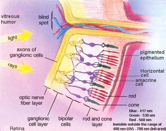
Figure 12b Retina |
reach the photoreceptors, incoming light must first pass through all the other layers of cells in the retina. There are five layers altogether (see Figure 12b). Starting from the outermost layer: |
 |
|
Figure 13 Optic Pathway |
 |
 |
According to the frequency of the sound wave, different parts of the basilar membrane along the organ of Corti are set into motion. In general, low-pitch sounds make the apex of the cochlea vibrate while high-pitched ones cause most vibrations near the base of the cochlea. Figure 15 shows such frequency distribution along the length of the cochlea for both the incoming and outgoing waves. The strength of nerve signals also depends on the volume of the sound. This is interpreted by the brain as loudness. It is believed that tone is an interpretation of the brain based on the distribution of hair cells stimulated. |
Figure 14 Cochlea |
Figure 15 Sound Wave |
 |
|
Figure 16 Sense of Smell |
 |
|
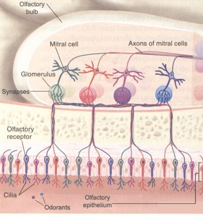
Figure 17 Olfactory Bulb |
 |
|
Figure 18 Pathways [view large image] |
The sense of taste and the sense of smell supplement each other, creating a combined effect when interpreted by the cerebral cortex. For example, some of the molecules may move from the nose down into the mouth region and stimulate the taste buds there. Therefore, part of what we refer to as smell actually may be taste. |
 |
 |
tongue (see Figure 19, also a 2010 version). Each detects a different class of chemical: sweet (sugars), sour (acids), bitter (complex organics), and salty (salts). The "hot" sensation of foods such as chili peppers is detected by pain receptors, not chemical receptors. But a report in 2006 reveals that contrary to popular belief, there is no tongue map. Responsiveness to the five basic modalities - bitter, sour, sweet salty and umami (a Japanese word meaning the savory or meaty taste of |
Figure 19 Tongue |
Figure 20 Papillae [view large image] |
amino acids) is present in all areas of the tongue. |
 |
|
Figure 21 Sense of Taste |
and starches. Likewise, taste is is extremely sensitive to bitter flavors, because many poisonous berries, fruits and fungi are bitter-tasting. |
 |
 |
receive the same attention of the brain. The relative importance is often represented by mapping over the length of the sensory or motor cortex. These cortical maps (Figure 22b) are not drawn to scale; instead they are variously distorted to reflect the amount the neural processing power devoted to different regions. This accounts for the grotesque appearance of the human body in the homun-culus, which is a translation of the body's sensory map into the human form. |
Figure 22a Propriocep-tors [view large image] |
Figure 22b Homunculus |
 |
|
Figure 23a Balance |
The vestibular nerve feeds its information chiefly to the cerebellum and to four structures in the medulla known as vestibular bodies. Using these data, as well as input from the other three sensory sources, the brain works out what to do, usually subconsciously. |
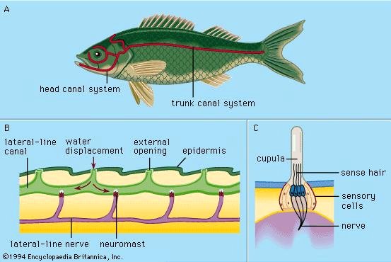 |
It turns out that such structure of hair within gel to detect disturbance has been around hundred of million years in the shark and fish (Figure 23b). This is the neuromasts embedded in the skin of fish. They give the fish information about the flow of water. Amphibians and reptiles have a simple uncoiled inner ear. Jawless fish has only one semicircular canal instead of three in mammals (for detecting three dimensional movement). Ultimately, it is the Pax 2 gene that give rise to these structures. It is also known that the Pax 6 gene is responsible for the development of eye. The connection to ancient creatures goes even deeper when it is |
Figure 23b Neuromast |
found that the box jellyfish carries a gene which is the combination of Pax 2 and Pax 6. The box jellyfish is an amazing animal with more than 20 eye pits and many eyes very similar to ours. They seem to double for ears as well. |