| Structure |
Location |
Functions |
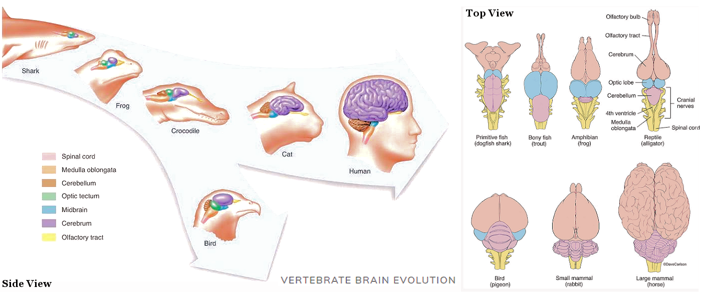
Hindbrain
(Reptilian Brain) |
|
|
| Medulla |
at the top of the spinal cord |
controls breathing, heart rate, and blood pressure. |
| Pons |
above the medulla |
regulates sensory information and facial expressions. |
| Cerebellum |
at the lower rear |
controls movement, coordination, balance, muscle tone, and learning motor skills. |
| Reticular Formation |
a network of nerves extends from the medulla to the cerebrum |
monitors the general level of activity in the hindbrain and maintains a state of arousal; essential for the regulation of sleep and wakefulness. |
| Midbrain (superior & inferior colliculus) |
above the pons between the hindbrain and forebrain |
relays sensory information from the spinal cord to the forebrain. |
| Pineal Gland |
on top of the midbrain behind the thalamus (the third eye¤ for fishes, amphibians, reptiles, and some birds) |
involves in circadian and circannual rhythms; possibly involves in maturation of sex organs. |
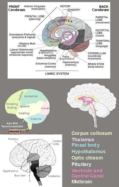
Limbic System
(Mammalian Brain) |
|
|
| Thalamus |
in the middle of the limbic system |
relays incoming information (except smell) to the appropriate part of the brain for further processing. |
| Hypothalamus, Pituitary Gland |
beneath thalamus |
regulates basic biological drives, hormonal levels, sexual behavior, and controls autonomic functions such as hunger, thirst, and body temperature. |
| Optic Chiasm |
in front of the pituitary gland |
left-right optic nerves cross-over point. |
| Septum |
adjacent to hypothalamus |
stimulates sexual pleasure |
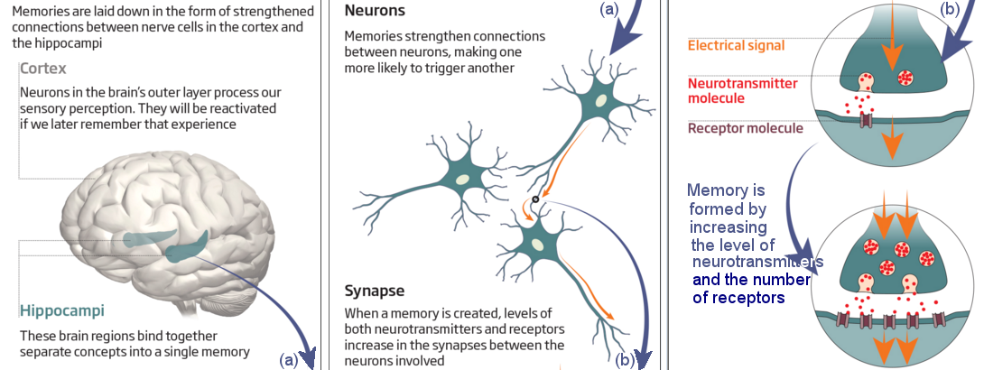
| Hippocampus |
within the temporal lobe |
mediates learning and memory formation. |
| Amygdala |
in front of the hippocampus |
responsible for anxiety, emotion, and fear |
| Mammillary Body, Fornix |
linked to the hippocampus |
have a role in emotional behavior, learning, and motivation. |
| Basal Ganglia (Striatum): Caudate Nucleus, Putamen, Globus Pallidus |
outside the thalamus |
involves in movement, emotions, planning and in integrating sensory information |
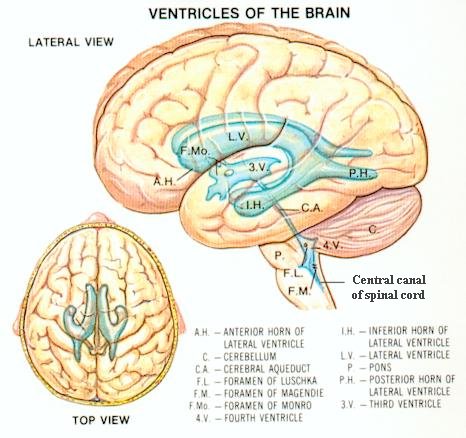
| Ventricles and Central Canal |
from tiny central canal within the spinal cord to the enlarged hollows within the skull called ventricles |
fills with cerebrospinal fluid for mechanical protection. |
| Cingulate Gyrus |
above corpus callosum |
concentrates attention on adverse internal stimuli such as pain, contains the feeling of self. |
| Corpus Callosum |
under the cingulate gyrus |
is a bundle of nerve fibers linking the cerebral hemispheres, involve in language learning. |
Forebrain
(Human Brain) |
|
|
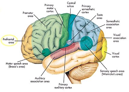
Frontal Lobe
(Conscious Brain) |
in front of the head |
controls voluntary movement, thinking, and feeling. |
| Prefrontal Cortex |
in front of the frontal lobe |
inhibits inappropriate actions, forms plans and concepts, helps focus attention, and bestows meaning to perceptions. |
| Parietal Lobe |
in top rear of the head |
contains the primary somatosensory area that manages skin sensation. |
| Occipital Lobe |
in the back of the head |
contains the visual cortex to manage vision. |
| Temporal Lobe |
on each side of the head above the temples |
contains the auditory cortex to manage hearing and speech. |
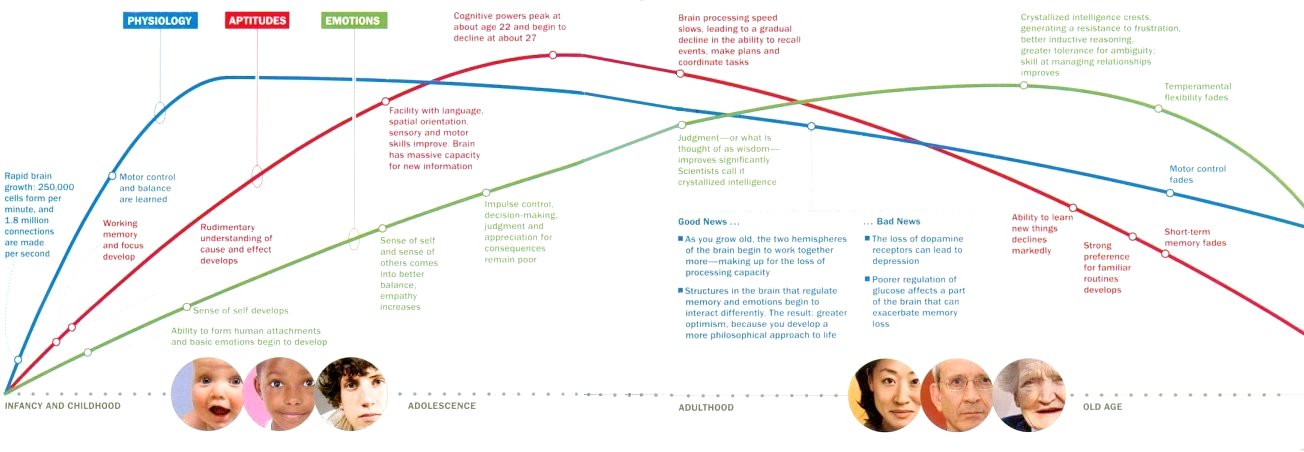
| Stage |
Age |
Event(s) |
DO |
DON'T |
| 1 |
0 - 10 months
Gestation |
* Growing neurons and connections
* Making sure each section of the brain grows properly and in the right place |
Mother should:
* be stress-free, eats well
* take folic acid and vitamin B12
* stimulate the young brain with sounds and sensations
|
* Mother should stay away from cigarettes, alcohol and other toxins
|
| 2 |
Birth - 6
Childhood |
* A sense of self develops as the parietal and frontal lobe circuits become more integrated.
* Development of voluntary movement, reasoning, and perception
* Frontal lobes become active leading to the development of emotions, attachments, planning, working memory and attention
* Life experiences shape the emotional well-being in adulthood
* At age 6, the brain is 95% of its adult weight and at its peak of energy consumption
|
* Parents should provide a nurturing environment and one-on-one interaction
|
* Parents should beware of the emotional consequence of neglect or harsh parenting
|
| 3 |
7 - 22
Adolescence |
* Wiring of the brain is still in progress
* Grey matter (neural connections) pruning
* White matter (fatty tissue surrounding neurons) increase helps to speed up electrical impulses and stabilize connections
* The prefrontal cortex (involving control of impulses, judgment and decision-making) is the last to mature
|
* Teenagers should learn to control reckless, irrational and irritable behaviors
* Do learn a skill to support life in the future
|
* Teenagers should avoid alcohol abuse, smoking, drug and unprotected sex.
|
| 4 |
23 - 65
Adulthood |
* The brain reaches the peak power at around age 22 and lasts for about 5 years; thereafter it's downhill all the way
* The last to mature and first to go brain functions are those involve executive control in the prefrontal and temporal cortices
* Episodic memory for recalling events also declines rapidly
* Processing speed slows down
* Working memory is able to store less information
|
* Stay active mentally and physically
* Eat healthy diet
|
* Avoid cigarettes, booze, and mind-altering drugs. |
| 5 |
> 65
Old Age |
* Losing brain cells in critical areas such as the hippocampus where memories are processed
|
* Exercise to improve abstract reasoning and concentration
* Learn new skill such as guitar playing to attain the same effect
* Practice meditation can promote neutral emotions
|
* Avoid grumpiness by eating certain foods, such as yogurt, chocolate, and almonds to get a good dose of dopamine (for promoting positive emotions)
* Don't stressed out as it is related to higher risk of developing dementia.
|
| Receptor |
Type |
Sense |
Stimulus / Limits |
| General |
|
|
|
| Ruffini's endings, Krause end bulbs |
Radioreceptor |
Hot-cold |
Heat flow |
| Merkel's and Meissner's endings |
Mechanoreceptor |
Touch |
Mechanical displacement of tissue |
| Pacinian corpuscles |
Mechanoreceptor |
Pressure |
Mechanical displacement of tissue |
| Free nerve endings |
Chemoreceptor |
Pain |
Tissue damage |
| Proprioceptors |
Mechanoreceptor |
Limb placement |
Mechanical displacement |
| Special |
|
|
|
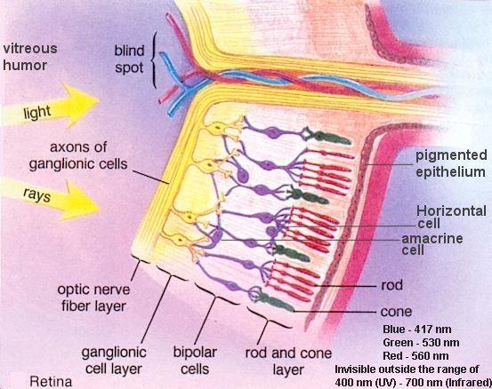
| Eye |
Radioreceptor |
Sight |
Light / wave of (370 - 730) nm, dimmest star at 10-14 watt/cm2 ~ 104 photons/sec-cm2,
and resolution of ~ 1' (arc minute) |
| Ear |
Mechanoreceptor |
Hearing |
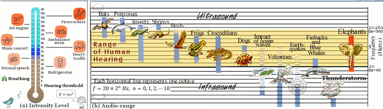
Sound / frequency range of (20 - 20K) Hz |
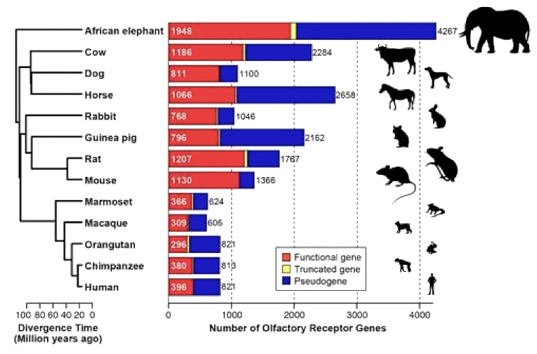
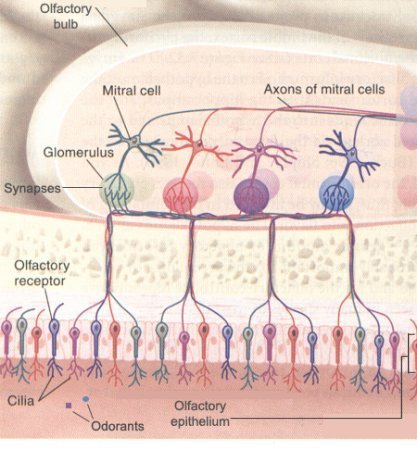
| Olfactory cells |
Chemoreceptor |
Smell |
Chemicals / 400 types of receptors, 109 different odours |
| Taste buds |
Chemoreceptor |
Taste |
Chemicals / 104 taste buds |
| Type |
Location(s) |
Function |
Example(s) |
| Working Memory |
|
|
|
| Phonological Loop |
Left hemisphere |
Rehearsing verbal information to keep it in the short-term memory |
String of numerals and alphabets such as telephone numbers |
| Visual-spatial Scratch Pad |
Visual Cortex |
Controlling visual imagery |
Scanning text |
| Central Executive |
Frontal lobe |
Controlling awareness of the information in working memory |
Constructing sentence, comprehending speech |
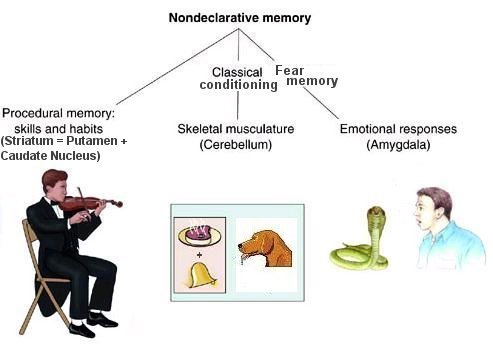
| Non-declarative Memory |
|
|
|
| Procedural Memory |
Cerebellum, temporal lobes |
Managing "how to" |
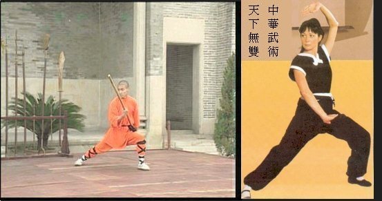
Riding a bicycle, kungfu exercise |
| Classical Conditioning |
Cerebellum |
Forming habitual behaviour |
Coffee break, afternoon tea |
| Fear Memory |
Amygdala |
Emotional conditioning |
Phobias, flashbacks |
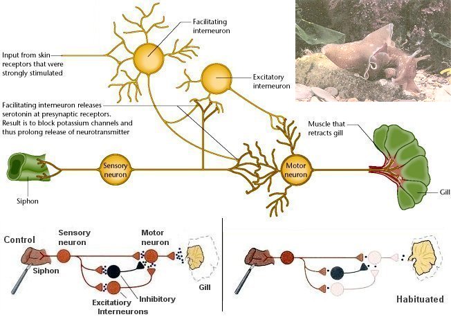
| Nonassociative Memory |
Spinal cord |
Habituation and Sensitization |
Decreased or increased responsiveness to stimulus |
| Remote Memory (Priming) |
Scattered around the cortex |
Foundation for new memories |
Childhood memory |
| Declarative Memory |
|
|
|
| Episodic Memory |
Cortex |
Remembering past experience |
Some enchanted evening |
| Semantic Memory |
Frontal lobe, temporal lobe |
Registering facts |
Meanings of words and symbols |


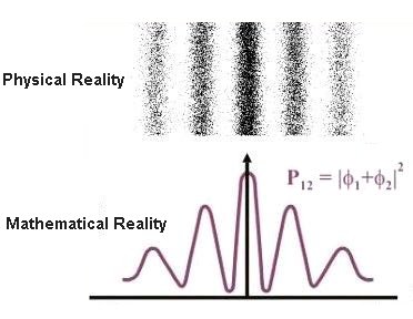
 = constant,
 )1/2 ~ 3.5x104 cm/sec (in air),
 R2), where R is the distance to the source, L the luminosity (intrinsic brightness = power)


 ) - "so of itself") and harmoniously.
 ) - "non-forcing" or "effortless action." It doesn't mean doing nothing, but rather acting in a way that flows with circumstances instead of fighting them.
 ); while sharing some core principles, Zen have distinct origins and approaches. Taoism, with its roots in ancient Chinese philosophy, emphasizes living in harmony with the Tao (often translated as "the Way"
  ), which is the unknowable reality of the universe. Zen Buddhism, on the other hand, originated in India and was significantly shaped by Chinese thought, particularly Taoism. It focuses on direct experience and insight into reality through meditation and mindfulness practices.